Knee Special Tests for Meniscus Tear: Procedures & Accuracy
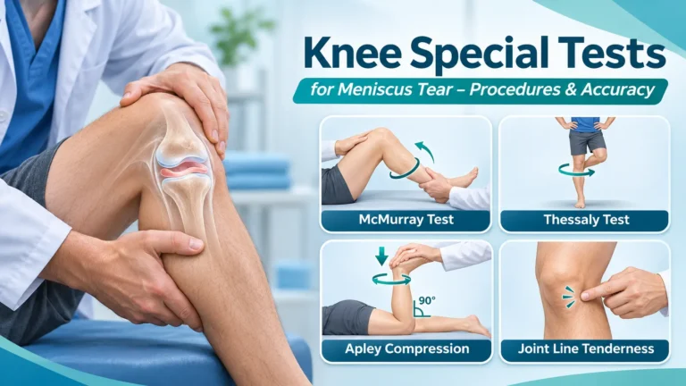
Knee special tests for meniscus tears include the McMurray test, Thessaly test, joint line tenderness assessment, and Apley compression test. These maneuvers help clinicians evaluate patients with knee pain, catching, swelling, or mechanical symptoms after twisting injuries, but no single test reliably confirms or excludes every meniscal tear.
This guide compares common meniscus examination tests, their procedures and positive findings, and how they fit into a safe clinical assessment. It also links to the detailed test articles already published on Special Tests & Examination.
Quick reference: special tests for meniscus tears
| Test | Procedure in brief | Suggestive findings / cautions |
|---|---|---|
| Joint line tenderness | Palpate the medial and lateral tibiofemoral joint lines | Focal tenderness; not specific to a tear |
| Medial McMurray test | Flex and extend the knee while applying tibial rotation and a valgus bias | Reproduction of joint-line pain or a palpable/audible click |
| Lateral McMurray test | Flex and extend with opposite tibial rotation and a varus bias | Lateral joint-line pain or mechanical click |
| Thessaly test | Supported weight-bearing pivot with the knee slightly flexed | Joint-line pain, catching, or locking; avoid if unsafe |
| Apley compression test | Prone, knee flexed to 90°, gentle axial compression and rotation | Joint-line pain; limited standalone accuracy |
| Ege test | Controlled squat with feet positioned according to the side tested | Pain or click during loaded flexion; not for acutely unstable knees |
| Bounce home test | Observe return toward knee extension from a flexed position | Pain or restricted terminal extension; nonspecific |
Key principle: A positive meniscus special test raises clinical suspicion but does not independently establish tear location, pattern, reparability, or the need for surgery.
What is a meniscus tear?
The knee has a medial and a lateral meniscus—fibrocartilaginous structures that distribute load, contribute to joint stability, and help absorb forces. A tear can follow an acute twisting or pivoting event, sometimes with a planted foot, or develop in association with age-related and degenerative tissue changes.
A traumatic meniscal injury may cause focal joint-line pain, swelling that develops over time, discomfort with squatting or twisting, and catching or locking. However, clicks are common in knees without a clinically significant tear, and degenerative meniscal changes can appear on MRI without being the main cause of symptoms. Clinical context matters.
Start with a safe knee examination
- History: identify twisting trauma, pivoting, sports participation, onset of swelling, giving way, persistent loss of extension, and previous knee problems.
- Safety screen: consider fracture, significant ligament injury, infection, or other urgent pathology. A painful hot swollen knee with fever, major deformity, neurovascular compromise, or inability to bear weight after significant trauma needs urgent assessment.
- Inspect and palpate: note swelling, effusion, bruising, patellar position, and medial or lateral joint-line tenderness.
- Range of motion: assess available extension and flexion; document a true mechanical extension block separately from pain-limited movement.
- Ligament screen: if the mechanism suggests associated instability, consider the Lachman test and appropriate collateral ligament tests.
- Targeted meniscus testing: select tests based on symptoms and tolerability. Do not provoke twisting loads in an acutely painful, unstable, or markedly swollen knee.
For the broader examination sequence, see our knee examination guide, knee range of motion, and knee effusion assessment.
1. Joint line tenderness test
With the patient sitting or supine and the knee comfortably flexed, palpate along the medial and lateral tibiofemoral joint lines. Ask whether palpation reproduces familiar pain and compare the other knee.
Positive finding: localized medial or lateral joint-line tenderness can support suspected meniscal injury, particularly after a convincing twisting mechanism. However, osteoarthritis, capsular irritation, and other conditions can produce similar tenderness. Joint-line palpation should not be interpreted alone.
2. McMurray test: medial and lateral meniscus
The McMurray test applies controlled knee flexion, rotation, and extension to provoke symptoms associated with meniscal pathology. Perform gently and stop with substantial pain or resistance.
Medial McMurray test
- Position the patient supine and flex the knee as comfortably tolerated, supporting the heel and knee.
- Externally rotate the tibia relative to the femur and apply a gentle valgus bias while gradually extending the knee.
- Palpate the medial joint line and ask about pain, catching, or a palpable click.
A reproduction of medial joint-line pain or a meaningful mechanical click may increase suspicion. Read the detailed medial McMurray test.
Lateral McMurray test
- Support the flexed knee and rotate the tibia internally.
- Apply a gentle varus bias while extending the knee within the patient’s comfortable range.
- Assess lateral joint-line symptoms and mechanical findings.
See the lateral McMurray test guide. Keep in mind that different standardized descriptions of McMurray testing exist and that technique, tear characteristics, and accompanying injuries influence results.
Interpretation: document the exact finding—pain, palpable click, restriction, or apprehension—rather than recording “McMurray positive” without context. Absence of symptoms does not rule out a tear.
3. Thessaly test
The Thessaly test evaluates pain or mechanical symptoms during controlled weight-bearing rotation. Because it requires pivoting, it should be reserved for a patient who can stand safely and tolerate the movement.
- Ask the patient to stand on the affected leg while holding the examiner’s hands or another stable support for balance.
- With the knee flexed approximately 20° (some protocols also use 5°), have the patient gently rotate the body and knee internally and externally in a controlled manner.
- Stop immediately if the knee feels unstable, locks, or causes significant pain.
- Document reproduction of joint-line pain, catching, or a locking sensation.
A positive Thessaly test can contribute to the overall probability of a meniscal tear, but accuracy in routine clinical populations varies. The maneuver is not appropriate for every acute knee injury. See the Thessaly test article.
4. Apley compression test
The Apley compression test is conducted with the patient prone and the knee flexed to approximately 90°.
- Stabilize the thigh and support the ankle.
- Apply gentle axial compression through the tibia toward the knee.
- Rotate the tibia internally and externally while asking where symptoms occur.
Positive result: joint-line pain with compression and rotation may support meniscal involvement. It can also provoke symptoms from other structures, so it is not a definitive meniscus test. See the Apley compression test. The related Apley distraction test is a distinct maneuver and is sometimes used to explore different sources of pain; neither should be treated as diagnostic on its own.
5. Ege test and bounce home test
Ege test
The Ege test uses a weight-bearing squat with foot rotation to provoke meniscal symptoms. Because it loads a flexed knee, reserve it for patients with adequate strength, comfort, and stability. Record joint-line pain or clicking rather than assuming that any squat discomfort indicates a tear. Read our Ege test guide.
Bounce home test
The bounce home test evaluates whether the knee returns smoothly toward full extension and whether pain or a mechanical restriction is present. Limited terminal extension may occur with several conditions, including swelling or a displaced meniscal fragment; it is not specific for a tear. See the bounce home test.
Which test is most accurate for a meniscus tear?
There is no universally most accurate bedside test. The American Academy of Orthopaedic Surgeons (AAOS) 2024 guideline for acute isolated meniscal pathology gives a moderate-strength recommendation that clinical examination—including joint-line tenderness, McMurray, and Thessaly—can help diagnose acute tears and may be more accurate when combined. Test performance varies by population, examiner, tear pattern, and associated injury.
The AAOS guideline applies specifically to acute isolated meniscal injuries. It should not automatically be extended to chronic degenerative tears, meniscal root tears, or knees with an accompanying ACL injury.
Meniscus tear vs ACL injury vs osteoarthritis
| Clinical feature | Considerations |
|---|---|
| Twisting with focal joint-line pain and delayed swelling | Can be consistent with traumatic meniscal injury |
| Pivoting injury, pop, rapid swelling, instability | Assess for ACL injury and other significant internal derangement |
| Gradual pain with stiffness and degenerative findings | Consider osteoarthritis and degenerative meniscal changes |
| Persistent inability to fully extend the knee | Evaluate urgently for a true mechanical block, including possible displaced meniscal tear |
These are clinical clues, not mutually exclusive diagnostic rules. For ACL assessment, see our Lachman test and knee anterior drawer test.
When is MRI or specialist referral appropriate?
According to the AAOS acute isolated meniscal pathology guideline, MRI is the preferred imaging modality for diagnosing suspected acute meniscal tears when imaging is indicated. Decisions depend on examination findings, severity, suspected associated damage, whether results would change management, and local pathways. An MRI finding must still be correlated with symptoms and physical examination, particularly in older patients with common age-related meniscal changes.
A truly locked knee (a persistent mechanical block to extension), major traumatic swelling or suspected multi-structure injury, significant instability, concerning neurovascular findings, or a hot febrile joint should not be managed by repeated provocative testing alone. Arrange timely or urgent assessment based on severity.
Step-by-step clinical examination checklist
- Document injury mechanism, timing, symptoms, locking versus clicking, and weight-bearing tolerance.
- Screen for urgent conditions and consider established fracture imaging criteria where applicable.
- Check effusion, knee extension and flexion, and both joint lines.
- Assess ligament stability where clinically indicated.
- Use McMurray and/or Thessaly selectively when the patient can tolerate them.
- Add Apley or other maneuvers only if they provide useful additional information.
- Record findings, the most likely diagnosis and differentials, and an imaging or referral plan when needed.
Frequently asked questions
What are the three common special tests for a meniscus tear?
Joint-line tenderness, McMurray, and Thessaly are the examinations emphasized by the 2024 AAOS acute isolated meniscal pathology guideline.
Which test is best for the medial meniscus?
Medial joint-line palpation and a medial McMurray maneuver are common choices. The best approach is to combine findings with the mechanism, symptoms, range of motion, and assessment for other injuries.
Can a negative McMurray test rule out a meniscus tear?
No. A tear may be present despite a negative McMurray test, especially when the knee is painful, guarded, or has a tear pattern that is not readily provoked.
Should the Thessaly test be used immediately after injury?
Not automatically. It is a weight-bearing twisting maneuver that may be inappropriate in a painful, unstable, markedly swollen, or non-weight-bearing knee. Safety and clinical need take priority.
Does every meniscus tear require surgery?
No. Management depends on the type and location of the tear, symptoms, locking, activity goals, age, associated injury, and response to appropriate nonoperative care. Treatment choices should be individualized by a qualified clinician.
Key takeaways
The McMurray, Thessaly, joint-line tenderness, and Apley tests are valuable as parts of a structured knee examination. Their main purpose is to refine clinical suspicion—not to replace history, fracture and ligament screening, or appropriate imaging and referral decisions. Avoid provocative weight-bearing maneuvers when the acute knee is unstable or cannot safely tolerate them.
References
- American Academy of Orthopaedic Surgeons. Acute Isolated Meniscal Pathology: Clinical Practice Guideline. Published June 10, 2024.
- AAOS. Guideline summary and diagnostic recommendations for acute isolated meniscal pathology. 2024.
- Brophy RH, et al. Academy of Orthopaedic Surgeons Clinical Practice Guideline Summary: Management of Acute Isolated Meniscal Pathology. J Am Acad Orthop Surg. 2025.
- American Physical Therapy Association. AAOS Acute Isolated Meniscal Pathology guideline resource.