Standing Flexion Test (Piedallu Test): Procedure, Positive Results & Accuracy
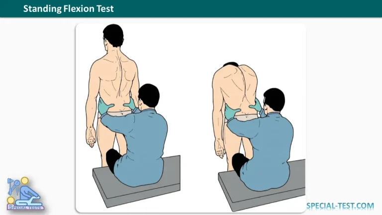
The Standing Flexion Test, also known as the Piedallu Test or Standing Forward Flexion Test, is a physical examination technique traditionally used to assess sacroiliac joint (SIJ) mobility and identify asymmetrical movement of the pelvis during forward bending.
Healthcare professionals, including orthopedic clinicians, physiotherapists, and osteopathic practitioners, use this test as part of a comprehensive examination of patients with lower back pain, pelvic discomfort, or suspected sacroiliac joint dysfunction.
The test involves palpating the posterior superior iliac spines (PSIS) while the patient bends forward. Differences in the movement of these bony landmarks may suggest altered lumbopelvic mechanics. However, research indicates that the Standing Flexion Test has limitations in reliability and diagnostic validity and should not be used independently to diagnose sacroiliac joint pathology.
What Is the Purpose of the Standing Flexion Test?
The primary purpose of the Standing Flexion Test is to evaluate asymmetry in pelvic movement during forward trunk flexion.
Traditionally, clinicians use the examination to:
- Assess movement patterns involving the sacroiliac joints and pelvis.
- Identify asymmetrical movement of the posterior superior iliac spines.
- Screen for possible sacroiliac joint mobility restrictions.
- Evaluate lumbopelvic movement in patients with lower back or pelvic pain.
- Guide further physical examination and clinical assessment.
The Standing Flexion Test is classified as a mobility or motion-palpation test, rather than a pain-provocation test. Its primary observation is the relative position and movement of pelvic landmarks, not whether the maneuver reproduces pain.
How to Perform the Standing Flexion Test
The Standing Flexion Test is performed with the patient standing while the examiner monitors the movement of both posterior superior iliac spines.
Patient Position
The patient stands upright with their back facing the examiner, both feet resting flat on the floor, and the knees extended.
The patient should maintain a comfortable standing position with weight distributed as evenly as possible between both lower limbs.
Examiner Position
The examiner stands or sits behind the patient and identifies the right and left posterior superior iliac spines.
Both thumbs are placed over these landmarks to compare their position and movement during forward bending.
Step-by-Step Procedure
- Identify the bony landmarks: Palpate both posterior superior iliac spines and compare their initial positions.
- Position the thumbs: Place one thumb over each PSIS, maintaining symmetrical contact without applying excessive pressure.
- Ask the patient to bend forward: Instruct the patient to slowly flex the trunk toward the floor while keeping both feet on the ground and the knees extended.
- Observe pelvic movement: Monitor whether one PSIS appears to move upward earlier or farther than the opposite PSIS.
- Compare both sides: Identify any asymmetry in the timing or relative displacement of the pelvic landmarks.
- Record the findings: Document the side demonstrating greater relative superior movement and any associated symptoms.
The examination should be performed slowly to allow the clinician to observe pelvic movement accurately.
What Is a Positive Standing Flexion Test?
A positive Standing Flexion Test is traditionally identified when one posterior superior iliac spine moves superiorly earlier or farther than the PSIS on the opposite side during forward trunk flexion.
According to the traditional biomechanical interpretation, the side showing greater upward displacement is considered the side with possible sacroiliac joint restriction or hypomobility.
For example:
- Positive right Standing Flexion Test: The right PSIS appears to move higher or earlier than the left.
- Positive left Standing Flexion Test: The left PSIS demonstrates greater relative superior movement.
- Negative Standing Flexion Test: Both PSIS landmarks show relatively symmetrical movement during forward bending.
Importantly, a positive finding does not necessarily indicate a mechanically blocked sacroiliac joint. Pelvic asymmetry may also result from differences in hip motion, muscle flexibility, pelvic anatomy, or lumbopelvic coordination.
Biomechanical Explanation of the Standing Flexion Test
The sacroiliac joints connect the sacrum to the right and left iliac bones. These joints provide stability and permit a small amount of movement during activities such as walking, standing, and bending.
One movement associated with the sacroiliac joint is nutation, in which the upper portion of the sacrum moves anteriorly and inferiorly relative to the ilia. The opposite movement is called counternutation.
Traditional explanations of the Standing Flexion Test suggest that restricted sacroiliac mobility may alter the movement of the corresponding ilium, producing asymmetrical PSIS displacement during trunk flexion.
However, actual sacroiliac joint movement is very small, and the displacement observed during the test cannot be attributed exclusively to movement within the SI joint.
A more contemporary interpretation considers the test an assessment of overall lumbopelvic movement rather than a direct measurement of sacroiliac joint motion.
Factors That Can Affect Standing Flexion Test Results
Several anatomical and biomechanical factors can influence the outcome of the Standing Flexion Test.
1. Hamstring tightness
Reduced hamstring flexibility may restrict pelvic rotation during forward bending and produce an altered movement pattern. Bilateral hamstring tightness may affect movement on both sides.
2. Leg-length discrepancy
Unequal lower limb lengths can create pelvic obliquity and influence the initial position of the posterior superior iliac spines. Clinicians should account for relevant leg-length differences when interpreting the test.
3. Hip mobility restrictions
Limited hip flexion or asymmetrical hip range of motion may cause compensatory pelvic movement and contribute to an apparently positive finding.
4. Lumbar spine stiffness
Reduced lumbar flexibility can modify the relationship between spinal and pelvic movements during trunk flexion.
5. Anatomical pelvic asymmetry
Natural differences in pelvic shape or bony landmark position may affect palpation and the interpretation of PSIS displacement.
For these reasons, the examiner should consider the patient’s overall musculoskeletal presentation rather than interpreting isolated PSIS asymmetry as confirmation of sacroiliac pathology.
Reliability and Diagnostic Accuracy of the Standing Flexion Test
Although the Standing Flexion Test is commonly taught in manual therapy and musculoskeletal examination, scientific evidence has raised concerns about its reliability and diagnostic accuracy.
A study by Vincent-Smith and Gibbons (1999) reported only 42% agreement between different examiners, with a kappa coefficient of 0.052. Agreement when the same examiner repeated the assessment was higher, at 68%.
A 2021 study by Ribeiro and colleagues found limited inter-rater reliability, despite observing some construct validity when the test was performed on healthy participants.
Furthermore, a systematic review and meta-analysis published in 2021 concluded that sacroiliac mobility palpation tests have inconsistent reliability and insufficient evidence supporting their diagnostic validity.
A more recent critical review published in 2026 emphasized that asymmetrical PSIS movement may reflect overall lumbopelvic mechanics rather than isolated sacroiliac joint dysfunction.
These findings indicate that a positive Standing Flexion Test cannot independently confirm sacroiliac joint dysfunction or establish the sacroiliac joint as the source of pain.
Clinical Significance
The Standing Flexion Test may provide observational information about pelvic and trunk movement during a musculoskeletal examination.
Nevertheless, clinicians should interpret the findings alongside the patient’s medical history, pain distribution, functional limitations, neurological examination, and other relevant orthopedic tests.
When sacroiliac joint pain is suspected, additional tests may include:
- FABER (Patrick’s) Test: Evaluates hip and pelvic pain reproduction during flexion, abduction, and external rotation.
- Thigh Thrust Test: Applies a posteriorly directed force through the femur to provoke sacroiliac region pain.
- Sacral Thrust Test: Applies pressure over the sacrum to assess whether familiar pain is reproduced.
- SI Joint Compression Test: Applies compressive force across the pelvis to assess pain provocation.
- SI Joint Distraction Test: Applies an anteriorly directed separating force to the pelvic bones.
Research suggests that combinations of pain-provocation tests can provide more useful diagnostic information than isolated mobility tests, although even positive test clusters cannot definitively confirm sacroiliac joint pain.
Frequently Asked Questions (FAQs)
1. What does the Standing Flexion Test assess?
The Standing Flexion Test assesses asymmetrical pelvic movement during forward trunk bending. It is traditionally used to screen for possible sacroiliac joint mobility restrictions, but it does not directly measure SI joint motion.
2. What indicates a positive Standing Flexion Test?
A positive test occurs when one posterior superior iliac spine appears to move upward earlier or farther than the opposite side. Traditionally, this finding is associated with possible restriction on the side demonstrating greater relative movement.
3. Is the Standing Flexion Test reliable?
Research has demonstrated variable reliability, particularly when different examiners perform the test. Although examiner training and standardized techniques may improve agreement, its diagnostic accuracy remains uncertain.
4. Can the Standing Flexion Test diagnose sacroiliac joint dysfunction?
No. The test alone cannot reliably establish sacroiliac joint dysfunction or determine whether the SI joint is responsible for a patient’s pain. A comprehensive clinical assessment is necessary.
5. What is the difference between the Standing Flexion Test and the Seated Flexion Test?
The Standing Flexion Test evaluates pelvic landmark movement while the patient bends forward from a standing position. The Seated Flexion Test uses a similar palpation technique while the patient is sitting. Sitting reduces some influences from the lower limbs, although neither test independently confirms SI joint pathology.
6. Can tight hamstrings cause a positive Standing Flexion Test?
Yes. Reduced hamstring flexibility can influence pelvic motion during forward bending and may produce asymmetrical findings. Therefore, clinicians should consider hamstring flexibility and hip mobility when interpreting the results.
7. Is the Standing Flexion Test a pain-provocation test?
No. It is primarily a motion-palpation test used to observe asymmetrical pelvic movement. Pain-provocation tests, such as the thigh thrust and sacral thrust tests, evaluate whether specific mechanical stresses reproduce familiar symptoms.
Conclusion
The Standing Flexion Test (Piedallu Test) is a commonly described physical examination maneuver for assessing pelvic movement and traditionally screening for sacroiliac joint dysfunction.
A positive result is characterized by asymmetrical superior displacement of the posterior superior iliac spines during forward trunk flexion. Historically, this finding has been interpreted as possible restriction of the sacroiliac joint on the affected side.
However, scientific evidence indicates that the test has limited diagnostic validity and inconsistent inter-examiner reliability. Pelvic asymmetry may reflect several musculoskeletal factors rather than isolated sacroiliac joint dysfunction.
Consequently, the Standing Flexion Test should be considered a supplementary assessment technique and interpreted within a comprehensive clinical examination rather than used as a standalone diagnostic tool.
References
- Orthofixar. Standing Flexion Test: Procedure, Interpretation, and Clinical Accuracy. https://orthofixar.com/special-test/standing-flexion-test/
- Vincent-Smith B, Gibbons P. Inter-examiner and intra-examiner reliability of the standing flexion test. Manual Therapy. 1999;4(2):87–93. PubMed – PMID: 10509062
- Ribeiro RP, et al. Construct validity and reliability of tests for sacroiliac dysfunction: standing flexion test (STFT) and sitting flexion test (SIFT). Journal of Osteopathic Medicine. 2021;121(11):849–856. PubMed – PMID: 34551460
- Validity and Reliability of Palpatory Clinical Tests of Sacroiliac Joint Mobility: A Systematic Review and Meta-analysis. 2021. PubMed – PMID: 33896601
- The Standing Forward Flexion Test in Manual Therapy: A Critical Review and a Functional Reinterpretation. 2026. NCBI PubMed Central
- Dreyfuss P, et al. Positive sacroiliac screening tests in asymptomatic adults. Spine. 1994. PubMed – PMID: 8059269
- Sacroiliac Joint Pain. StatPearls. NCBI Bookshelf. https://www.ncbi.nlm.nih.gov/books/NBK470299/
- Diagnostic Accuracy of Clusters of Pain Provocation Tests for Detecting Sacroiliac Joint Pain: Systematic Review With Meta-analysis. Journal of Orthopaedic & Sports Physical Therapy. 2021. PubMed – PMID: 34210160