Prone Knee Flexion Test: Purpose, Procedure, Positive Findings & Accuracy
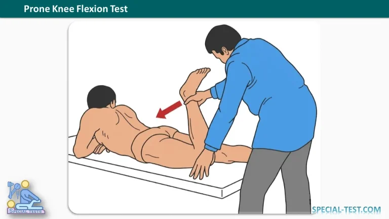
The Prone Knee Flexion Test is a clinical orthopedic examination used to help differentiate pain originating from the lumbar spine and sacroiliac (SI) joint. It may also provide information about lumbar nerve root irritation, femoral nerve sensitivity, and tightness of the quadriceps muscles.
This test is commonly performed during the physical examination of patients presenting with low back pain, sacroiliac joint dysfunction, or suspected lumbar radiculopathy.
Although the Prone Knee Flexion Test can provide useful clinical findings, it should be interpreted alongside the patient’s medical history, neurological examination, and other orthopedic special tests rather than used as a standalone diagnostic procedure.
What Is the Purpose of the Prone Knee Flexion Test?
The primary purpose of the Prone Knee Flexion Test for the lumbar spine is to assist clinicians in distinguishing between lumbar spine pain and sacroiliac joint-related pain.
The test may be useful when evaluating patients with suspected:
- Sacroiliac joint pain or pelvic ligament dysfunction.
- Lumbar spine disorders involving the intervertebral discs.
- Upper lumbar nerve root irritation, particularly involving L2, L3, or L4.
- Femoral nerve irritation or increased neural mechanosensitivity.
- Quadriceps muscle tightness or injury.
- Mechanical low back pain associated with altered lumbopelvic movement.
Passive knee flexion stretches the anterior thigh structures and may influence the position of the pelvis and lumbar spine. These mechanical effects can reproduce symptoms originating from different tissues.
Consequently, the location and nature of the patient’s symptoms are important when interpreting the results.
How to Perform the Prone Knee Flexion Test
The Prone Knee Flexion Test is performed with the patient lying face down on an examination table.
Patient Position: Prone lying.
Examiner Position: Standing beside the lower extremity being examined.
Step-by-Step Procedure
- Position the patient: Ask the patient to lie comfortably in the prone position with both lower extremities extended and relaxed.
- Stabilize the pelvis: Observe and, when appropriate, stabilize the pelvis to minimize excessive rotation or compensatory lumbar movement.
- Flex the knee passively: Grasp the patient’s lower leg or ankle and slowly bend the knee, bringing the heel toward the buttock as far as comfortably possible.
- Observe the patient’s response: During passive knee flexion, monitor for pain, muscular tightness, neurological symptoms, or compensatory pelvic movement.
- Apply resistance: Ask the patient to attempt knee extension against the examiner’s resistance while controlling the movement. This resisted component is included in the traditional lumbar Prone Knee Flexion Test description.
- Compare both sides: Repeat the examination on the opposite leg and document differences in symptoms, flexibility, and movement.
The examiner should avoid forcing the knee into excessive flexion or continuing the examination when severe pain or significant neurological symptoms occur.
What Is a Positive Prone Knee Flexion Test?
Traditionally, the Prone Knee Flexion Test is considered positive when knee flexion produces abnormal tension or pain involving the sacroiliac joint, lumbosacral junction, or lumbar spine.
Classical descriptions suggest that tension may be experienced sequentially in the sacroiliac region, lumbosacral junction, and lumbar spine.
However, this sequence has not been established as a reliable diagnostic criterion for identifying the precise anatomical source of pain.
A clinically meaningful finding is the reproduction of the patient’s familiar symptoms during the test.
Interpretation of Positive Test Findings
1. Sacroiliac Joint Pain
Pain localized around the sacroiliac joint or posterior pelvis may suggest SI joint involvement.
Potential contributing factors include ligamentous irritation, degenerative changes, or altered pelvic biomechanics.
Nevertheless, pain in this region is not specific to SI joint pathology. Additional sacroiliac provocation tests are required to strengthen the clinical assessment.
2. Lumbar or Lumbosacral Pain
Pain reproduced in the lower lumbar region without radiating neurological symptoms may suggest a mechanical source of low back pain.
Possible structures involved include the intervertebral discs, facet joints, muscles, and supporting ligaments.
The Prone Knee Flexion Test alone cannot reliably distinguish these individual structures.
3. Radiating Pain and Lumbar Nerve Root Irritation
Reproduction or worsening of radiating leg pain may raise suspicion of lumbar nerve root involvement.
The femoral nerve stretch component is particularly relevant to the L2, L3, and L4 nerve roots.
Upper lumbar radiculopathy commonly produces symptoms in the groin, anterior thigh, or medial thigh rather than following the typical posterior leg distribution associated with lower lumbar radiculopathy.
Additional examination findings, such as muscle weakness, sensory changes, and altered patellar reflexes, can help support suspected nerve root involvement.
4. Anterior Thigh Pain or Paresthesia
Pain, tingling, or abnormal sensations in the anterior thigh during knee flexion may indicate:
- Femoral nerve irritation.
- Upper lumbar nerve root involvement.
- Rectus femoris or quadriceps muscle tightness.
- Quadriceps muscle injury.
- Other hip or anterior thigh musculoskeletal disorders.
Anterior thigh discomfort alone does not confirm femoral neuropathy or lumbar disc herniation.
Prone Knee Flexion Test Accuracy and Reliability
The diagnostic accuracy of the Prone Knee Flexion Test as an isolated examination remains insufficiently established.
Cibulka and Koldehoff (1999) investigated a cluster of four sacroiliac joint tests involving 219 participants with and without low back pain.
The combined testing approach demonstrated:
- Sensitivity: 82%
- Specificity: 88%
- Positive Predictive Value: 86%
- Negative Predictive Value: 84%
Importantly, these values apply to the combined cluster of sacroiliac joint tests, not the isolated Prone Knee Flexion Test.
Furthermore, a 2021 systematic review and meta-analysis concluded that positive clusters of sacroiliac pain provocation tests have limited ability to definitively identify the SI joint as the source of pain. Negative clusters may be more useful for reducing suspicion of sacroiliac joint pain.
Research on femoral nerve stretch testing suggests potential usefulness in detecting midlumbar nerve root involvement, although diagnostic performance varies between studies.
Therefore, Prone Knee Flexion Test findings should always be integrated with the overall clinical examination.
Prone Knee Flexion Test vs. Femoral Nerve Stretch Test
The Prone Knee Flexion Test and Femoral Nerve Stretch Test share similar examination movements, but their clinical emphasis can differ.
The traditional lumbar Prone Knee Flexion Test is described as an assessment of pain associated with the lumbar spine and sacroiliac region, sometimes including resisted knee extension.
The Femoral Nerve Stretch Test, also called the Prone Knee Bend Test, primarily evaluates possible irritation of the upper lumbar nerve roots and femoral nerve.
During femoral nerve stretch testing, the examiner flexes the patient’s knee while the patient lies prone. Hip extension may be added to increase tension on the femoral nerve.
Reproduction of familiar radiating anterior thigh pain is more suggestive of neural involvement than an isolated sensation of muscular stretching.
Because these procedures overlap, clinicians should clearly document the technique used and the symptoms reproduced.
Clinical Limitations and Precautions
The Prone Knee Flexion Test has several important limitations.
First, knee flexion affects multiple anatomical structures simultaneously, including the quadriceps, hip, pelvis, lumbar spine, and neural tissues. This reduces the specificity of the examination.
Second, a positive result does not independently establish a diagnosis of disc herniation, sacroiliac ligament insufficiency, or nerve root compression.
Third, patients with significant knee pathology, acute injury, or severe pain may not tolerate the examination.
Clinical assessment should include neurological testing, lumbar movement assessment, hip examination, and appropriate sacroiliac provocation tests when indicated.
Patients with progressive neurological weakness, new bladder or bowel dysfunction, or saddle anesthesia require urgent medical evaluation rather than further provocative testing.
Frequently Asked Questions (FAQ)
1. What is the Prone Knee Flexion Test used for?
The Prone Knee Flexion Test is used to assist in evaluating lumbar spine pain, sacroiliac joint-related symptoms, and possible neural irritation. It helps identify movements that reproduce the patient’s familiar symptoms but cannot independently establish a definitive diagnosis.
2. What indicates a positive Prone Knee Flexion Test?
A positive finding generally involves reproduction of the patient’s familiar lumbar, sacroiliac, or radiating leg pain during prone knee flexion. The clinical significance depends on the symptom distribution and the exact examination technique.
3. Can the Prone Knee Flexion Test detect a herniated lumbar disc?
The test may reproduce symptoms associated with lumbar nerve root irritation, including irritation caused by disc herniation. However, a positive test cannot confirm disc herniation. A clinical neurological examination and, when indicated, MRI are needed for further assessment.
4. Which nerve roots are assessed during prone knee bending?
The femoral nerve stretch component primarily assesses the upper and middle lumbar nerve roots, especially L2, L3, and L4. Irritation involving these roots may produce radiating pain or abnormal sensations in the anterior or medial thigh.
5. Is the Prone Knee Flexion Test the same as the Femoral Nerve Stretch Test?
Not exactly. The movements overlap, and the names are sometimes used interchangeably. However, the traditional Prone Knee Flexion Test described for lumbar and sacroiliac pain may include resisted knee extension, whereas the Femoral Nerve Stretch Test primarily evaluates neural symptom reproduction during knee flexion, sometimes combined with hip extension.
6. Can the Prone Knee Flexion Test diagnose sacroiliac joint dysfunction?
No. Pain near the sacroiliac joint during the examination may raise suspicion, but it is not sufficient to diagnose SI joint dysfunction. A combination of clinical history, multiple SI joint provocation tests, and selected confirmatory investigations provides a more reliable assessment.
7. What causes anterior thigh pain during the Prone Knee Flexion Test?
Anterior thigh pain can result from tight or injured quadriceps muscles, stretching of the rectus femoris, femoral nerve irritation, or upper lumbar radiculopathy. The examiner must differentiate muscular stretching discomfort from the reproduction of familiar neurological symptoms.
Conclusion
The Prone Knee Flexion Test is a useful component of the orthopedic examination for patients presenting with lumbar spine pain, sacroiliac joint symptoms, or possible upper lumbar nerve root irritation.
The examination involves passive knee flexion while the patient lies prone, with an additional resisted knee extension maneuver included in the traditional lumbar test.
Pain in the sacroiliac, lumbar, or anterior thigh regions may provide clues about the underlying disorder. However, these findings are not sufficiently specific to identify a single pathological structure.
For accurate clinical interpretation, the Prone Knee Flexion Test should be combined with neurological assessment, other orthopedic special tests, and appropriate diagnostic investigations.
References
- OrthoFixar. Prone Knee Flexion Test: Differentiate Lumbar Spine vs. Sacroiliac Pain. https://orthofixar.com/special-test/prone-knee-flexion-test/
- Cibulka MT, Koldehoff R. Clinical usefulness of a cluster of sacroiliac joint tests in patients with and without low back pain. J Orthop Sports Phys Ther. 1999;29(2):83–92. PubMed – PMID: 10322583
- Herron LD, Pheasant HC. Prone knee-flexion provocative testing for lumbar disc protrusion. Spine. 1980;5(1):65–67. PubMed – PMID: 6444765
- Saueressig T, et al. Diagnostic Accuracy of Clusters of Pain Provocation Tests for Detecting Sacroiliac Joint Pain: Systematic Review With Meta-analysis. J Orthop Sports Phys Ther. 2021;51(9):422–431. PubMed – PMID: 34210160
- Suri P, et al. The accuracy of the physical examination for the diagnosis of midlumbar and low lumbar nerve root impingement. Spine. 2011. PubMed – PMID: 20543768
- OrthoFixar. Femoral Nerve Stretch Test: Diagnosing High Lumbar Radiculopathy. https://orthofixar.com/special-test/femoral-nerve-stretch-test/
- NCBI Bookshelf. Sacroiliac Joint Pain. StatPearls. https://www.ncbi.nlm.nih.gov/books/NBK470299/
- OrthoFixar. Prone Knee Bending Test: Screening for Upper Lumbar Radiculopathy. https://orthofixar.com/special-test/prone-knee-bending-test/