Triceps Reflex Test: C7–C8 Nerve Roots, Grading & Technique
What Is the Triceps Reflex Test?
The triceps reflex test, also known as the triceps tendon reflex or triceps jerk reflex, is a neurological examination used to evaluate the integrity of the C7–C8 cervical nerve roots, radial nerve, and associated spinal cord reflex pathways.
It is one of the commonly assessed deep tendon reflexes (DTRs) during an upper limb neurological examination. Healthcare professionals use this test to identify potential abnormalities involving peripheral nerves, cervical nerve roots, and the central nervous system.
The test involves striking the distal triceps tendon with a reflex hammer, producing an involuntary contraction of the triceps brachii muscle and subsequent elbow extension.
The triceps reflex is particularly useful when assessing patients with suspected cervical radiculopathy, radial nerve dysfunction, or cervical myelopathy. However, its findings must always be interpreted alongside muscle strength, sensation, and other neurological examination results.
Triceps Reflex Nerve Root and Anatomy
The triceps reflex primarily assesses the C7 nerve root, with contributions from C8 and, in some anatomical descriptions, C6.
The main anatomical structures involved include:
- Nerve roots: C7–C8, predominantly C7.
- Peripheral nerve: Radial nerve.
- Muscle tested: Triceps brachii.
- Tendon tested: Distal triceps tendon, immediately proximal to the olecranon.
- Expected movement: Involuntary elbow extension.
How Does the Triceps Reflex Work?
The triceps reflex is a muscle stretch reflex mediated by a neurological pathway known as the reflex arc.
When the examiner taps the triceps tendon, the resulting brief stretch activates muscle spindles within the triceps brachii. Sensory impulses travel through the radial nerve toward the cervical spinal cord.
The spinal cord then activates motor neurons that transmit impulses back through the radial nerve, causing the triceps muscle to contract and the elbow to extend.
An intact reflex suggests that the tested sensory and motor pathways are functioning, although it does not exclude all neurological disorders.
How to Perform the Triceps Reflex Test
Correct patient positioning, muscle relaxation, and reflex hammer technique are essential for obtaining reliable results.
1. Patient Position
The patient should sit comfortably, preferably looking away from the tested arm.
The upper limb must remain relaxed to prevent voluntary muscle contraction from interfering with the reflex response.
2. Examiner Position
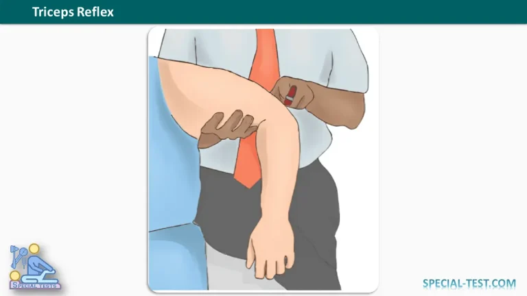
The examiner stands beside the patient and supports the upper limb.
One commonly used technique involves:
- Abducting the patient’s shoulder to approximately 90 degrees.
- Flexing the elbow to approximately 90 degrees.
- Supporting the arm to allow the triceps muscle to relax.
- Keeping the forearm free to move during reflex testing.
Alternatively, the arm may be supported in a comfortable, partially flexed position.
3. Identify the Triceps Tendon
Locate the distal triceps tendon on the posterior aspect of the elbow, immediately above its insertion into the olecranon process of the ulna.
4. Tap the Tendon
Using a neurological reflex hammer, deliver a quick, controlled tap directly over the triceps tendon.
The strike should be sufficiently brisk to produce a stretch response without causing unnecessary discomfort.
5. Observe the Response
A normal response consists of:
Involuntary contraction of the triceps brachii muscle, producing a brief extension movement at the elbow.
The examiner should assess the strength of the response and compare it with the opposite arm.
If the reflex is difficult to elicit, verify that the muscle is relaxed, adjust the limb position, and repeat the examination.
Triceps Reflex Grading: 0 to 4+
The triceps reflex is commonly assessed using a standardized deep tendon reflex grading scale.
| Grade | Reflex Response | Clinical Interpretation |
|---|---|---|
| 0 | No reflex elicited | Absent reflex (areflexia) |
| 1+ | Reduced or barely detectable response, sometimes requiring reinforcement | Hyporeflexia |
| 2+ | Normal reflex response | Normal reflex activity |
| 3+ | Brisker-than-average response | Brisk reflex; may be normal or abnormal |
| 4+ | Markedly increased reflex activity, potentially accompanied by clonus | Marked hyperreflexia |
Important: Reflex grades should be evaluated in context. A symmetrical 1+ or 3+ response can be normal in some individuals. By contrast, new or unexplained reflex asymmetry may indicate an underlying neurological problem.
What Does an Abnormal Triceps Reflex Indicate?
An abnormal triceps reflex may suggest dysfunction involving the cervical nerve roots, peripheral nerves, spinal cord, or descending motor pathways.
Absent or Reduced Triceps Reflex (Hyporeflexia)
A diminished or absent triceps reflex may occur with:
- C7 cervical radiculopathy.
- Certain C8 nerve root lesions.
- Radial nerve injury affecting the reflex pathway.
- Peripheral neuropathy.
- Lower motor neuron dysfunction.
A reduced triceps reflex is a potentially useful finding in patients presenting with neck pain, radiating arm pain, or weakness during elbow extension.
However, an absent reflex alone cannot confirm cervical radiculopathy.
Increased Triceps Reflex (Hyperreflexia)
An exaggerated triceps reflex may suggest dysfunction of upper motor neuron pathways.
Possible associated conditions include:
- Cervical spinal cord compression.
- Cervical myelopathy.
- Other central nervous system disorders affecting descending motor control.
An isolated brisk response is not necessarily pathological. Clinicians should look for additional findings such as increased muscle tone, pathological reflexes, or abnormal gait.
Inverted Triceps Reflex
An inverted or paradoxical triceps reflex occurs when tapping the triceps tendon produces elbow flexion rather than the expected elbow extension.
This unusual response may occur with cervical spinal cord or nerve root pathology, particularly when impairment of the triceps reflex arc coexists with increased activity in other muscle groups.
Clinical Importance of the Triceps Reflex Test
The triceps reflex examination is a useful component of the neurological assessment of the upper extremity.
Its main clinical applications include evaluating possible C7 nerve root dysfunction, examining radial nerve reflex pathways, identifying asymmetrical deep tendon reflexes, and detecting findings that may suggest upper or lower motor neuron involvement.
Research published in PubMed has identified reduced tendon reflexes as potentially specific clinical findings for cervical radiculopathy, although sensitivity and diagnostic performance vary between studies.
For this reason, the triceps reflex should be combined with other clinical assessments, including manual muscle testing, sensory examination, and additional neurological tests.
Depending on the patient’s symptoms and examination findings, further investigations such as magnetic resonance imaging or electrodiagnostic studies may be appropriate.
Frequently Asked Questions (FAQs)
1. What nerve root is tested by the triceps reflex?
The triceps reflex mainly tests the C7 nerve root, with contributions from C8. Some anatomical references also describe C6 involvement. The reflex travels through the radial nerve.
2. What is the normal response to the triceps reflex test?
The normal response is contraction of the triceps brachii muscle followed by a brief elbow extension. A reflex grade of 2+ is generally considered normal.
3. Which nerve innervates the triceps reflex?
The radial nerve provides the peripheral innervation involved in the triceps tendon reflex. The reflex pathway primarily involves the C7–C8 cervical spinal segments.
4. What does an absent triceps reflex mean?
An absent triceps reflex may indicate dysfunction involving the C7–C8 nerve roots, radial nerve, or another component of the reflex arc. The finding must be compared with the opposite arm and interpreted with other neurological signs.
5. What does a brisk triceps reflex indicate?
A brisk triceps reflex may represent normal individual variation or increased reflex activity associated with an upper motor neuron disorder. Reflex asymmetry and accompanying neurological abnormalities are important when interpreting this result.
6. Can the triceps reflex identify cervical radiculopathy?
The triceps reflex can help identify neurological deficits associated with cervical radiculopathy, particularly C7 nerve root involvement. However, it cannot independently establish the diagnosis. A comprehensive neurological examination is necessary.
7. What is the difference between the biceps and triceps reflexes?
The biceps reflex primarily evaluates the C5–C6 nerve roots through the musculocutaneous nerve and produces elbow flexion. In comparison, the triceps reflex primarily evaluates C7–C8 through the radial nerve and produces elbow extension.
8. Why should the triceps reflex be tested on both arms?
Comparing the right and left triceps reflexes helps identify asymmetry. A significant difference between sides may be more clinically meaningful than a mildly diminished or brisk reflex that is symmetrical.
Conclusion
The triceps reflex test is a simple but important neurological examination for assessing the C7–C8 nerve roots, radial nerve, and triceps brachii reflex pathway.
A normal response produces involuntary elbow extension, while reduced, absent, or exaggerated reflex activity may provide clues to peripheral or central neurological dysfunction.
Accurate technique, standardized reflex grading, and comparison between both upper limbs are essential for clinical interpretation. The triceps reflex is most informative when combined with a complete neurological assessment rather than used as an isolated diagnostic test.
References
- Orthofixar. Triceps Reflex Test: Technique, Innervation, and Reflex Grading. https://orthofixar.com/special-test/triceps-reflex-test/
- Orthofixar. Deep Tendon Reflex Testing: Examination Technique and Grading. https://orthofixar.com/special-test/deep-tendon-reflex-testing/
- NCBI Bookshelf – StatPearls. Physiology, Deep Tendon Reflexes. https://www.ncbi.nlm.nih.gov/books/NBK562238/
- Walker HK. Deep Tendon Reflexes. Clinical Methods: The History, Physical, and Laboratory Examinations. 3rd edition. NCBI Bookshelf. https://www.ncbi.nlm.nih.gov/books/NBK396/
- NCBI Bookshelf – StatPearls. Neuroanatomy, Spinal Cord Myotatic Reflex. https://www.ncbi.nlm.nih.gov/books/NBK551629/
- Yousif MS, et al. (2025). Neurological Examination for Cervical Radiculopathy: A Scoping Review. BMC Musculoskeletal Disorders. https://pubmed.ncbi.nlm.nih.gov/40188056/
- Nonoperative Management of Cervical Radiculopathy. American Family Physician, 2016. PubMed. https://pubmed.ncbi.nlm.nih.gov/27175952/