Seated Flexion Test: Procedure, Positive Findings, SI Joint Dysfunction & Diagnostic Accuracy
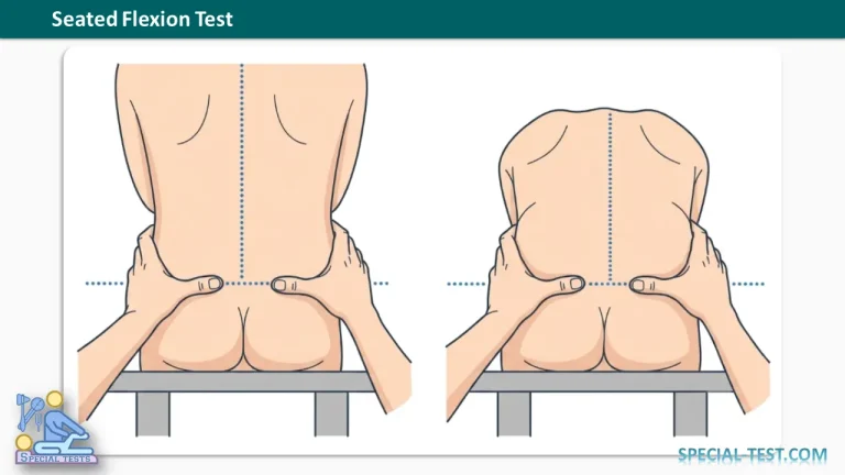
The Seated Flexion Test, also known as the Sitting Flexion Test or Seated Forward Flexion Test, is a physical examination technique traditionally used to assess sacroiliac joint (SI joint) dysfunction and asymmetrical pelvic movement.
This orthopedic assessment is commonly performed in osteopathic medicine, physical therapy, and manual therapy. During the examination, the clinician palpates the posterior superior iliac spines (PSIS) while the patient bends forward from a seated position.
The test aims to detect differences in pelvic landmark movement that may suggest restricted sacroiliac mobility. However, scientific evidence indicates that the Seated Flexion Test has limited diagnostic accuracy when used independently and should not be considered a definitive test for SI joint pathology.
What Is the Purpose of the Seated Flexion Test?
The primary purpose of the Seated Flexion Test is to evaluate possible sacroiliac joint movement restriction by assessing the symmetry of posterior pelvic landmark motion.
The sacroiliac joints connect the sacrum to the iliac bones of the pelvis. These joints provide stability and transmit forces between the spine and lower extremities while allowing a small amount of movement.
Clinicians traditionally use the Seated Flexion Test to assess:
- Possible sacroiliac joint hypomobility or dysfunction.
- Asymmetrical movement of the posterior pelvis.
- Functional restrictions involving the sacrum and iliac bones.
- Pelvic movement patterns during forward trunk flexion.
Unlike the Standing Flexion Test, the seated position reduces some influences from the lower extremities. Traditionally, this was believed to help isolate movement around the sacroiliac joints. However, sitting does not completely eliminate contributions from the lumbar spine, hips, surrounding muscles, or soft tissues.
How to Perform the Seated Flexion Test
The Seated Flexion Test is performed with the patient sitting and the examiner positioned behind them.
Patient Position
- Ask the patient to sit upright on a firm examination table.
- Position the patient with the legs hanging over the table edge and the feet comfortably supported.
- Ensure the patient maintains a relatively symmetrical sitting posture.
- The examiner stands directly behind the patient to observe the posterior pelvic landmarks.
Examiner Position and Procedure
- Identify the PSIS landmarks: Palpate the left and right posterior superior iliac spines.
- Position the thumbs: Place each thumb immediately inferior (caudal) to the corresponding PSIS.
- Maintain bilateral contact: Keep both landmarks under simultaneous observation and palpation.
- Instruct the patient to bend forward: Ask the patient to slowly flex the trunk forward at the waist as far as comfortably possible.
- Observe pelvic movement: Compare the timing and superior displacement of both PSIS landmarks.
- Evaluate movement symmetry: Determine whether both sides move similarly or whether one PSIS appears to rise earlier or farther than the other.
The examination should be performed gently, without forcing the patient into painful trunk flexion.
What Is a Positive Seated Flexion Test?
Interpretation of the Seated Flexion Test is based on the relative superior movement of the left and right PSIS during forward bending.
Normal (Negative) Seated Flexion Test
A negative test is traditionally characterized by:
- Both PSIS landmarks moving superiorly during trunk flexion.
- Relatively symmetrical movement between the left and right sides.
- No obvious difference in the timing or extent of PSIS elevation.
These findings suggest symmetrical pelvic landmark movement during the examination, although they do not exclude sacroiliac joint dysfunction or pain.
Positive Seated Flexion Test
A positive test occurs when one PSIS moves superiorly earlier or farther than the opposite PSIS.
Traditionally, the side demonstrating greater superior movement is interpreted as the side of possible sacroiliac restriction or pelvic fixation.
For example, if the right PSIS rises farther than the left during forward trunk flexion, the examination is traditionally interpreted as positive on the right side.
However, this asymmetry does not establish that the right sacroiliac joint is mechanically restricted. Differences in pelvic anatomy, muscle tension, lumbar movement, and palpation technique can influence the result.
Seated Flexion Test Sensitivity and Specificity
The diagnostic accuracy of the Seated Flexion Test has been investigated in clinical research.
A 1999 study by Levangie examined several sacroiliac joint mobility tests, including the sitting forward flexion test, and their association with measured innominate torsion.
The following values have been reported for the Seated Flexion Test:
| Diagnostic measure | Reported value |
|---|---|
| Sensitivity | 9% |
| Specificity | 93% |
What Do These Results Mean?
Sensitivity of 9%: The test identified only a small proportion of individuals classified as having innominate torsion according to the study’s reference measurements. This indicates a high likelihood of false-negative findings for that outcome.
Specificity of 93%: The test was negative in a high proportion of individuals without the measured condition. Although high specificity can sometimes help support a diagnosis after a positive result, the overall diagnostic usefulness depends on the likelihood ratios and the quality of the reference standard.
Importantly, these values relate to identifying innominate torsion, not directly diagnosing painful sacroiliac joint dysfunction.
In Levangie’s study, the findings did not support the clinical usefulness of these tests for identifying innominate torsion. Therefore, the reported sensitivity and specificity should not be interpreted as proof that the Seated Flexion Test can reliably diagnose or exclude SI joint pain.
Reliability and Clinical Evidence
The reliability of the Seated Flexion Test remains an important limitation.
A 2021 systematic review and meta-analysis by Ribeiro and colleagues investigated the reliability and validity of palpatory sacroiliac joint mobility tests. The review reported a pooled intra-examiner kappa value of 0.68 for the sitting flexion test, suggesting relatively good agreement when the same examiner repeated the assessment.
However, the review did not identify studies establishing concurrent validity for these mobility tests.
Another 2021 study evaluated the sitting and standing flexion tests in 30 participants. For the Sitting Flexion Test, researchers reported:
- Construct validity: 56.7% agreement (κ = 0.29).
- Intra-examiner reliability: 56.7% agreement (κ = 0.38).
- Inter-examiner reliability: 13.3% agreement (κ = 0.01).
These findings highlight that results may differ considerably between examiners and that repeated palpation does not necessarily provide an accurate diagnosis of sacroiliac pathology.
Clinical Significance of the Seated Flexion Test
Although the Seated Flexion Test remains part of some traditional musculoskeletal examinations, it should be interpreted cautiously.
A comprehensive assessment of suspected sacroiliac joint pain should include the patient’s history, symptoms, physical examination findings, and consideration of alternative causes of lower back or pelvic pain.
Clinicians may use additional SI joint pain provocation tests, including:
- Thigh Thrust Test
- Sacroiliac Compression Test
- Sacroiliac Distraction Test
- Gaenslen’s Test
- FABER (Patrick’s) Test
These tests assess whether mechanical loading reproduces the patient’s familiar pain rather than attempting to identify small differences in sacroiliac mobility.
Research and clinical guidelines support interpreting multiple findings together rather than relying on a single examination maneuver. Even combinations of pain provocation tests have diagnostic limitations and cannot always confirm the sacroiliac joint as the source of pain.
Frequently Asked Questions (FAQ)
1. What is the Seated Flexion Test used for?
The Seated Flexion Test is traditionally used to assess suspected sacroiliac joint dysfunction by comparing movement of the posterior superior iliac spines during seated forward bending.
2. What indicates a positive Seated Flexion Test?
A positive result occurs when one PSIS moves superiorly earlier or farther than the opposite side. The side showing greater upward movement is traditionally considered the restricted side, although this interpretation has limited diagnostic validation.
3. What is the difference between the Seated Flexion Test and the Standing Flexion Test?
The main difference is patient positioning. The Seated Flexion Test is performed while sitting, whereas the Standing Flexion Test is performed while standing. Sitting reduces some influences from lower-extremity positioning but does not fully isolate sacroiliac joint movement.
4. What are the sensitivity and specificity of the Seated Flexion Test?
Reported values are approximately 9% sensitivity and 93% specificity in a study evaluating associations with innominate torsion. These figures should not be generalized to diagnosing sacroiliac joint pain.
5. Is the Seated Flexion Test reliable?
Research has produced mixed reliability findings. Some studies demonstrate reasonable repeatability within the same examiner, while others report poor agreement between different examiners. The test’s diagnostic validity remains uncertain.
6. Can the Seated Flexion Test confirm sacroiliac joint dysfunction?
No. A positive Seated Flexion Test alone cannot confirm sacroiliac joint dysfunction or identify the sacroiliac joint as the source of pain. Its findings must be considered alongside the clinical history and other appropriate assessments.
7. Does a negative Seated Flexion Test rule out SI joint dysfunction?
No. The low reported sensitivity means that a negative result does not reliably exclude the condition being assessed. A normal test also cannot rule out sacroiliac joint pain.
Key Takeaways
The Seated Flexion Test is a traditional orthopedic mobility examination used to compare posterior pelvic landmark movement during seated trunk flexion.
A positive finding consists of asymmetrical superior movement of the PSIS landmarks. Although this has traditionally been associated with sacroiliac joint restriction, available research does not establish that the finding reliably identifies joint pathology.
The commonly reported sensitivity of 9% and specificity of 93% relate to an investigation of innominate torsion, rather than a validated diagnosis of sacroiliac joint pain.
Consequently, the Seated Flexion Test should not be used as an isolated diagnostic tool. A more evidence-informed assessment incorporates the patient’s symptoms, relevant clinical examination findings, and appropriate supplementary investigations when indicated.
References
- OrthoFixar. Seated Flexion Test for SI Joint Dysfunction: Procedure, Positive Findings, and Diagnostic Accuracy. https://orthofixar.com/special-test/seated-flexion-test/
- Levangie PK. (1999). Four clinical tests of sacroiliac joint dysfunction: The association of test results with innominate torsion among patients with and without low back pain. Physical Therapy, 79(11), 1043–1057. PubMed – PMID: 10534797
- Ribeiro RP, et al. (2021). Validity and reliability of palpatory clinical tests of sacroiliac joint mobility: A systematic review and meta-analysis. Journal of Manipulative and Physiological Therapeutics, 44(4), 307–318. PubMed – PMID: 33896601
- Ribeiro RP, et al. (2021). Construct validity and reliability of tests for sacroiliac dysfunction: Standing flexion test (STFT) and sitting flexion test (SIFT). Journal of Osteopathic Medicine, 121(11), 849–856. PubMed – PMID: 34551460
- Klerx SP, et al. (2020). Clinimetric properties of sacroiliac joint mobility tests: A systematic review. Musculoskeletal Science and Practice. PubMed – PMID: 31744776
- Saueressig T, et al. (2021). Diagnostic accuracy of clusters of pain provocation tests for detecting sacroiliac joint pain: Systematic review with meta-analysis. Journal of Orthopaedic & Sports Physical Therapy, 51(9), 422–431. [PubMed – PMID: 34210160](https://pubmed.ncbi.nlm.nih.gov