Achilles Tendon Reflex Test: How to Perform, S1 Nerve Root Assessment & Interpretation
What Is the Achilles Tendon Reflex?
The Achilles tendon reflex, also known as the ankle jerk reflex or S1 deep tendon reflex, is a neurological examination used to assess the integrity of the S1 nerve root, tibial nerve, and associated spinal reflex pathways.
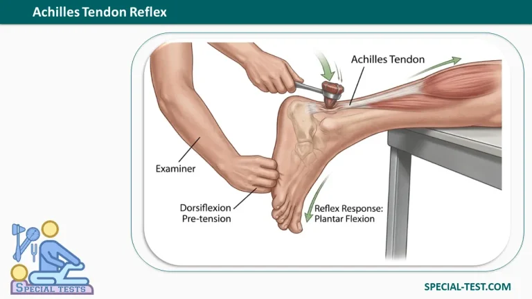
The test involves tapping the Achilles tendon with a reflex hammer to produce an involuntary contraction of the calf muscles, resulting in plantar flexion of the foot (downward movement at the ankle).
The Achilles reflex is an essential component of the lower extremity neurological examination. It helps clinicians identify potential abnormalities involving the peripheral nerves, spinal nerve roots, and central nervous system.
The test is particularly useful when evaluating suspected S1 radiculopathy, peripheral neuropathy, and upper or lower motor neuron dysfunction.
What Does the Achilles Tendon Reflex Test Assess?
The Achilles tendon reflex primarily evaluates the S1 nerve root, with additional contributions from S2.
The main anatomical structures involved include:
| Component | Anatomical Structure |
|---|---|
| Reflex name | Achilles tendon reflex (ankle jerk) |
| Primary nerve root | S1 |
| Secondary nerve root | S2 |
| Peripheral nerve | Tibial nerve |
| Muscles tested | Gastrocnemius and soleus (triceps surae) |
| Tendon | Achilles tendon (calcaneal tendon) |
| Normal response | Plantar flexion of the foot |
| Reflex classification | Deep tendon (myotatic) reflex |
Physiology of the Achilles Reflex Arc
The Achilles reflex is a muscle stretch reflex that operates through a spinal reflex arc.
When the Achilles tendon is tapped, the gastrocnemius and soleus muscles are briefly stretched. This activates muscle spindles, which transmit sensory signals through the tibial nerve to the spinal cord, primarily at the S1 level.
The spinal cord subsequently sends motor impulses back through the tibial nerve, producing contraction of the calf muscles and plantar flexion of the ankle.
An intact reflex indicates that the involved sensory and motor pathways are functioning sufficiently to produce a response.
How to Perform the Achilles Tendon Reflex Test
The Achilles tendon reflex test is performed using a neurological reflex hammer. Correct patient positioning, adequate muscle relaxation, and appropriate tendon tension are necessary for reliable results.
Patient Position
The patient is commonly positioned prone on an examination table, with both feet extending beyond the edge.
Alternatively, the patient may be examined while seated with the legs relaxed or lying supine with the knee slightly flexed.
Examiner Position
The examiner stands or sits beside the patient and supports the foot near its neutral position, applying slight passive dorsiflexion to place the Achilles tendon under appropriate tension.
Step-by-Step Examination Procedure
- Position the patient: Ask the patient to lie prone with the feet extending beyond the examination table.
- Relax the muscles: Ensure the gastrocnemius and soleus muscles are relaxed.
- Support the foot: Hold the foot and apply slight passive dorsiflexion without excessive stretching.
- Locate the Achilles tendon: Identify the tendon immediately above its insertion on the posterior calcaneus.
- Apply the stimulus: Tap the Achilles tendon briskly using a reflex hammer.
- Observe the response: Look for contraction of the calf muscles and plantar flexion of the foot.
- Compare both sides: Repeat the examination on the opposite leg to identify asymmetry.
The tendon should not be excessively stretched or left completely slack, as either condition may interfere with eliciting the reflex.
What If the Achilles Reflex Cannot Be Elicited?
When the reflex is difficult to obtain, the examiner should first verify positioning, muscle relaxation, and correct hammer technique.
A reinforcement technique known as the Jendrassik maneuver may help. The patient interlocks the fingers of both hands and attempts to pull them apart while the examiner repeats the tendon tap.
An alternative plantar tap technique may also be useful, particularly in older patients or individuals with limited mobility.
Achilles Tendon Reflex Grading and Interpretation
The Achilles reflex is commonly graded using a standardized 0 to 4+ deep tendon reflex scale.
| Grade | Reflex Response | Clinical Interpretation |
|---|---|---|
| 0 | No reflex elicited | Absent reflex (areflexia) |
| 1+ | Reduced or weak reflex, sometimes elicited only with reinforcement | Hyporeflexia |
| 2+ | Normal reflex response | Normal |
| 3+ | Brisk reflex response | Upper range of normal or increased reflex activity |
| 4+ | Markedly increased reflex, potentially accompanied by clonus | Hyperreflexia |
Important: A 3+ reflex is not necessarily pathological, particularly when reflexes are symmetrical. A 4+ response may be accompanied by clonus, but clonus is not required under every grading convention.
What Is the Normal Achilles Tendon Reflex Response?
A normal response consists of a visible or palpable contraction of the gastrocnemius and soleus muscles, producing plantar flexion of the ankle.
A 2+ reflex is conventionally documented as normal. However, interpretation depends on the patient’s age, baseline reflexes, examination technique, and comparison with the opposite limb.
What Does an Absent Achilles Reflex Indicate?
An absent Achilles tendon reflex (grade 0) may indicate dysfunction affecting the S1 nerve root, tibial nerve, or peripheral nervous system.
Possible causes include:
- S1 radiculopathy: Compression or irritation of the S1 nerve root, potentially associated with lumbar disc herniation.
- Peripheral neuropathy: Damage to peripheral nerves, including neuropathy associated with diabetes mellitus.
- Tibial nerve dysfunction: Injury or disease affecting the peripheral nerve responsible for the reflex.
- Other neurological or metabolic disorders: Conditions affecting reflex pathway function, including hypothyroidism.
Nevertheless, an absent Achilles reflex does not independently establish a diagnosis. Reflexes may be difficult to elicit in healthy individuals, particularly older adults.
What Does an Increased Achilles Reflex Indicate?
An exaggerated Achilles reflex may suggest upper motor neuron dysfunction, especially when associated with increased muscle tone, weakness, pathological reflexes, or clonus.
Hyperreflexia may occur in disorders affecting descending motor pathways, including spinal cord lesions.
The reflex finding must be interpreted together with the complete neurological examination.
Clinical Significance of the Achilles Tendon Reflex
The Achilles tendon reflex is particularly important when examining patients with lower back pain, sciatica, lower limb weakness, sensory disturbances, or suspected neurological disease.
A diminished or absent ankle jerk on one side may support a diagnosis of S1 nerve root dysfunction, particularly when accompanied by characteristic clinical findings such as plantar flexion weakness and sensory changes involving the lateral foot.
Bilateral reduction in Achilles reflexes may prompt investigation of peripheral neuropathy, depending on the patient’s history and additional examination findings.
Reflex testing is not sufficiently sensitive or specific to diagnose these conditions on its own. A normal reflex does not exclude radiculopathy or peripheral nerve disease.
Frequently Asked Questions (FAQ)
1. Which nerve root is tested by the Achilles tendon reflex?
The Achilles tendon reflex primarily assesses the S1 spinal nerve root, with a contribution from S2. The sensory and motor components of the reflex arc travel through the tibial nerve.
2. Which nerve is responsible for the ankle jerk reflex?
The tibial nerve carries the sensory and motor impulses involved in the ankle jerk reflex. It supplies the gastrocnemius and soleus muscles responsible for plantar flexion.
3. What is a positive Achilles tendon reflex test?
A normal elicited Achilles reflex produces calf muscle contraction and plantar flexion of the foot. Unlike many orthopedic special tests, the Achilles reflex is not conventionally classified as simply positive or negative. Its response is graded and interpreted clinically.
4. What does a 0 Achilles reflex mean?
A grade 0 Achilles reflex means that no reflex response was elicited. It may be associated with S1 radiculopathy, peripheral neuropathy, or other neurological conditions. Proper technique and reinforcement should be considered before interpreting the finding.
5. Can the Achilles reflex be absent in healthy people?
Yes. Achilles reflexes may become less prominent with increasing age and can be difficult to elicit in otherwise healthy individuals. Comparison between the right and left legs, together with other neurological findings, is essential.
6. What is the difference between the Achilles reflex and patellar reflex?
The Achilles reflex primarily assesses the S1 nerve root through the tibial nerve and calf muscles. The patellar reflex primarily assesses L4, with contributions from L2 and L3, through the femoral nerve and quadriceps muscle.
Both tests are important components of lower extremity neurological assessment.
7. Is the Achilles reflex test used to diagnose an Achilles tendon rupture?
No. The Achilles tendon reflex evaluates neurological function rather than directly testing tendon continuity. The Thompson test (calf squeeze test) is commonly used when an Achilles tendon rupture is suspected.
Conclusion
The Achilles tendon reflex test is a simple and valuable neurological examination used primarily to assess S1 nerve root function and the integrity of the tibial nerve reflex pathway.
The test is performed by tapping the Achilles tendon with a reflex hammer and observing the resulting plantar flexion of the foot.
Correct technique, standardized reflex grading, and comparison between both lower limbs are important for accurate interpretation.
Although diminished, absent, or exaggerated reflexes may suggest neurological abnormalities, findings must always be considered alongside muscle strength, sensation, other deep tendon reflexes, and the patient’s clinical history.
References
- Figliuzzi A, Alvarez R, Al-Dhahir MA. Achilles Reflex. StatPearls. NCBI Bookshelf. Updated June 5, 2023.
https://www.ncbi.nlm.nih.gov/books/NBK459229/ - Achilles Reflex Test: How to Perform and Interpret the Ankle Jerk. Orthofixar.
https://orthofixar.com/special-test/achilles-reflex-test/ - Deep Tendon Reflex Testing: Grading, Technique & Nerve Root Guide. Orthofixar.
https://orthofixar.com/special-test/deep-tendon-reflex-testing/ - Physiology, Deep Tendon Reflexes. StatPearls. NCBI Bookshelf.
https://www.ncbi.nlm.nih.gov/books/NBK562238/ - Deep Tendon Reflexes. Clinical Methods. NCBI Bookshelf.
https://www.ncbi.nlm.nih.gov/books/NBK396/ - Stam J. Physical diagnostics—tendon reflexes. Nederlands Tijdschrift voor Geneeskunde. 1999;143(16):848–851. PMID: 10347654.
https://pubmed.ncbi.nlm.nih.gov/10347654/ - Does This Patient With Diabetes Have Large-Fiber Peripheral Neuropathy? PubMed. PMID: 20407062.
https://pubmed.ncbi.nlm.nih.gov/20407062/