Patellar Reflex Test (Knee-Jerk Reflex): Purpose, Procedure, Nerve Roots & Grading
What Is the Patellar Reflex Test?
The patellar reflex test, also known as the knee-jerk reflex test or quadriceps reflex, is a neurological examination used to assess the integrity of the femoral nerve and the L2–L4 spinal nerve roots, particularly L3 and L4.
This test is one of the most commonly performed deep tendon reflex (DTR) examinations. It evaluates the body’s involuntary response to a sudden stretch of the quadriceps femoris muscle.
When the patellar tendon is tapped with a reflex hammer, the quadriceps muscle contracts automatically, producing a brief extension of the knee. This response helps healthcare professionals evaluate the function of the peripheral nervous system, spinal cord reflex pathways, and descending motor pathways.
The patellar reflex is frequently assessed during neurological examinations, orthopedic evaluations, and investigations of lower limb weakness or suspected lumbar nerve root dysfunction.
What Is the Patellar Reflex Used For?
The primary purpose of the patellar tendon reflex test is to evaluate the neurological pathways responsible for knee extension.
The test assesses the following structures:
- Spinal nerve roots: L2–L4, with particular clinical emphasis on L3 and L4.
- Peripheral nerve: Femoral nerve.
- Muscle involved: Quadriceps femoris.
- Tendon stimulated: Patellar tendon, located immediately below the patella.
- Expected response: Involuntary quadriceps contraction causing knee extension.
The examination may help identify abnormalities associated with lumbar radiculopathy, femoral neuropathy, peripheral neuropathy, and certain central nervous system disorders.
However, the patellar reflex alone cannot establish a neurological diagnosis. Findings must be interpreted alongside muscle strength, sensation, other reflexes, and the patient’s clinical history.
Patellar Reflex Arc and Neuroanatomy
The patellar reflex is a monosynaptic stretch reflex, meaning that the main excitatory reflex pathway involves a direct connection between a sensory neuron and a motor neuron in the spinal cord.
The reflex arc involves the following sequence:
- Tendon stimulation: A reflex hammer strikes the patellar tendon, briefly stretching the quadriceps muscle.
- Sensory receptor activation: Muscle spindles within the quadriceps detect the sudden stretch.
- Afferent signal transmission: Sensory impulses travel through the femoral nerve toward the L2–L4 spinal cord segments.
- Spinal cord integration: Sensory neurons activate alpha motor neurons within the spinal cord.
- Efferent signal transmission: Motor impulses travel through the femoral nerve back to the quadriceps muscle.
- Motor response: The quadriceps contracts, producing knee extension.
This reflex occurs without requiring conscious control, although descending pathways from the brain influence reflex excitability.
How to Perform the Patellar Reflex Test
Correct patient positioning, muscle relaxation, and appropriate percussion technique are essential for reliable examination results.
Patient Position
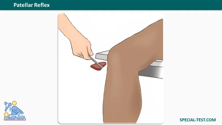
The patient should sit comfortably on an examination table with both knees flexed and the lower legs hanging freely over the edge.
The quadriceps muscles must remain relaxed throughout the examination. The patient may be instructed to look away from the tested leg to minimize anticipation.
If sitting is not possible, the test may be performed with the patient lying supine while the examiner supports the knee in slight flexion.
Examiner Position
The examiner stands or sits beside the patient, ensuring clear access to the anterior aspect of the knee.
The examiner should be positioned to observe the movement of the lower leg and, when necessary, palpate the quadriceps contraction.
Step-by-Step Examination Procedure
- Position the patient: Ask the patient to sit with the legs relaxed and hanging freely.
- Identify the patellar tendon: Locate the tendon immediately inferior to the patella and superior to the tibial tuberosity.
- Check muscle relaxation: Ensure the patient is not voluntarily contracting the quadriceps.
- Apply the stimulus: Using a reflex hammer, deliver a quick, controlled tap directly over the patellar tendon.
- Observe the response: Look for contraction of the quadriceps and a brief extension of the knee.
- Compare both sides: Repeat the examination on the opposite leg and assess any differences in reflex intensity.
- Document the findings: Record the reflex grade using the standard deep tendon reflex grading scale.
For patients with difficult-to-elicit reflexes, the examiner may use the Jendrassik maneuver, in which the patient hooks the fingers of both hands together and attempts to pull them apart while the tendon is tapped.
This reinforcement technique can increase reflex responsiveness and help identify a subtle response.
Patellar Reflex Test: Normal and Abnormal Results
What Is a Normal Patellar Reflex?
A normal patellar reflex produces a brief, involuntary contraction of the quadriceps femoris muscle accompanied by knee extension.
A 2+ reflex response is generally considered normal using the commonly applied clinical grading scale.
However, normal reflex intensity varies between individuals. Some healthy people demonstrate relatively brisk or diminished reflexes.
Symmetry between the two legs and consistency with other neurological findings are often more informative than the absolute reflex grade.
Patellar Reflex Grading Scale
| Grade | Reflex Response | Clinical Interpretation |
|---|---|---|
| 0 | No detectable reflex | Absent reflex (areflexia) |
| 1+ | Reduced or weak response, sometimes requiring reinforcement | Hyporeflexia |
| 2+ | Average reflex response | Normal |
| 3+ | Brisker-than-average response | May be normal or indicate hyperreflexia |
| 4+ | Markedly exaggerated response; clonus may be present | Significant hyperreflexia |
Important: Grading conventions vary slightly. Some systems specifically associate grade 4+ with clonus, while the NINDS grading system allows clonus to be documented separately. A brisk reflex is not automatically pathological.
Clinical Significance of Abnormal Patellar Reflexes
1. Diminished or Absent Patellar Reflex (0–1+)
A reduced or absent patellar tendon reflex may suggest dysfunction involving the lower motor neuron pathway or its associated reflex arc.
Possible causes include:
- Lumbar radiculopathy: Particularly involving the L3 or L4 nerve roots.
- Femoral neuropathy: Damage or compression affecting the femoral nerve.
- Peripheral neuropathy: Including neuropathy associated with diabetes mellitus.
- Spinal nerve root compression: Potentially caused by lumbar degenerative disease or intervertebral disc pathology.
- Neuromuscular disorders: Conditions affecting peripheral motor pathways or muscle function.
An absent reflex should not automatically be interpreted as a positive diagnostic finding. The examiner must consider reinforcement techniques, patient relaxation, bilateral comparison, and associated symptoms.
2. Exaggerated Patellar Reflex (3+–4+)
An abnormally increased patellar reflex may be associated with dysfunction of descending motor pathways, especially when accompanied by other upper motor neuron signs.
Potential clinical associations include:
- Spinal cord lesions affecting descending motor control.
- Stroke involving motor pathways.
- Multiple sclerosis.
- Other central nervous system disorders associated with upper motor neuron dysfunction.
Marked hyperreflexia may coexist with spasticity, increased muscle tone, or clonus.
Nevertheless, an isolated grade 3+ reflex may be a normal finding, particularly when responses are symmetrical.
3. Asymmetrical Patellar Reflexes
A significant difference between the right and left patellar reflexes may be more clinically meaningful than bilaterally brisk or reduced reflexes.
For example, a diminished right patellar reflex compared with the left may raise suspicion of right-sided femoral nerve dysfunction or L3–L4 radiculopathy.
Additional examinations, including quadriceps strength testing, sensory assessment, and other lower limb reflexes, are necessary to help localize the neurological abnormality.
Frequently Asked Questions (FAQs)
1. What nerve roots are tested by the patellar reflex?
The patellar reflex evaluates the L2–L4 spinal nerve roots, particularly L3 and L4. The L4 nerve root is often emphasized clinically when interpreting the knee-jerk reflex.
2. Which nerve controls the patellar reflex?
The femoral nerve carries both sensory and motor signals involved in the patellar reflex. It supplies the quadriceps femoris muscle, which produces knee extension.
3. What does a positive patellar reflex test mean?
The patellar reflex is generally documented by its grade rather than as simply positive or negative. A normal response involves quadriceps contraction and knee extension, typically graded 2+. Abnormal findings include absent, reduced, exaggerated, or asymmetrical responses.
4. What causes an absent knee-jerk reflex?
An absent patellar reflex may occur with femoral neuropathy, L3–L4 nerve root dysfunction, or peripheral neuropathy. Poor muscle relaxation or an inadequate testing technique may also make a reflex difficult to elicit.
5. What does an exaggerated patellar reflex indicate?
A markedly exaggerated patellar reflex can suggest upper motor neuron dysfunction, especially when associated with spasticity, clonus, or other abnormal neurological signs. However, a brisk reflex alone does not confirm neurological disease.
6. What is the normal grade for the patellar reflex?
A grade of 2+ is commonly considered normal. A grade of 1+ or 3+ may also occur in healthy individuals, depending on the clinical context and symmetry of reflex responses.
7. What is the Jendrassik maneuver?
The Jendrassik maneuver is a reinforcement technique used when deep tendon reflexes are difficult to elicit. The patient interlocks the fingers and attempts to pull the hands apart while the examiner taps the patellar tendon.
8. What is the difference between the patellar reflex and the Achilles reflex?
The patellar reflex tests the quadriceps muscle through the femoral nerve and L2–L4 spinal segments, whereas the Achilles reflex evaluates the calf muscles through the tibial nerve and predominantly the S1 nerve root, with S2 contribution.
Both examinations provide complementary information during a neurological assessment of the lower extremities.
Conclusion
The patellar reflex test is a quick, practical, and clinically valuable component of the neurological examination. It primarily assesses the integrity of the femoral nerve and L2–L4 spinal nerve roots, particularly L3 and L4.
A normal knee-jerk reflex involves involuntary quadriceps contraction and knee extension following percussion of the patellar tendon.
Diminished, absent, exaggerated, or asymmetrical reflex responses may help clinicians identify possible peripheral nerve, nerve root, spinal cord, or central motor pathway abnormalities.
For accurate interpretation, patellar reflex findings should always be evaluated in combination with the patient’s medical history and a comprehensive neurological examination.
References
- Orthofixar. Deep Tendon Reflex Testing: Grading, Technique & Nerve Root Guide. https://orthofixar.com/special-test/deep-tendon-reflex-testing/
- Orthofixar. Patellar Reflex Test: Clinical Significance, Grading, and Neurological Pathways. https://orthofixar.com/special-test/patellar-reflex-test/
- NCBI Bookshelf – StatPearls. Physiology, Deep Tendon Reflexes. https://www.ncbi.nlm.nih.gov/books/NBK562238/
- PubMed. Deep Tendon Reflexes. PMID: 30285397. https://pubmed.ncbi.nlm.nih.gov/30285397/
- PubMed Central (NCBI). Deep Tendon Reflex: The Tools and Techniques. What Surgical Neurology Residents Should Know. https://pmc.ncbi.nlm.nih.gov/articles/PMC8075597/
- PubMed. The Accuracy of Subjective Clinical Assessments of the Patellar Reflex. PMID: 23169260. https://pubmed.ncbi.nlm.nih.gov/23169260/