Jeanne’s Sign Test: Assessment of Ulnar Nerve Motor Weakness
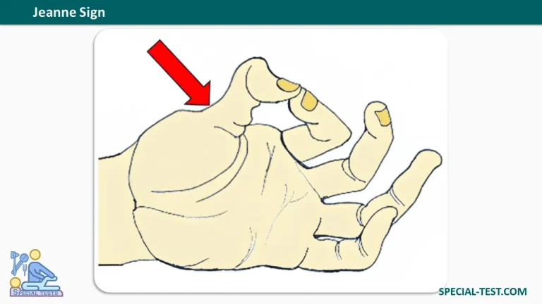
Jeanne’s Sign is a clinical examination finding used to assess ulnar nerve motor weakness, particularly weakness of the adductor pollicis muscle in the thumb. A positive Jeanne’s Sign is characterized by hyperextension of the thumb metacarpophalangeal (MCP) joint during key pinch.
This sign helps healthcare professionals identify abnormal thumb mechanics associated with ulnar nerve dysfunction. It is commonly assessed alongside Froment’s Sign and other clinical tests during the examination of patients with suspected ulnar nerve injury or compression.
What Is Jeanne’s Sign?
Jeanne’s Sign is an abnormal compensatory movement of the thumb observed when a patient performs a key pinch, also called a lateral pinch.
During normal key pinch, the thumb presses against the lateral surface of the index finger. This movement requires coordinated muscle activity, particularly the adductor pollicis, which is innervated by the deep branch of the ulnar nerve.
When the adductor pollicis becomes weak or paralyzed, the patient may compensate by hyperextending the thumb MCP joint to maintain pinch stability.
This abnormal movement is known as Jeanne’s Sign and may indicate an underlying ulnar nerve motor deficit.
What Is Jeanne’s Sign Used For?
The primary purpose of Jeanne’s Sign is to evaluate ulnar nerve motor dysfunction affecting thumb adduction and pinch strength.
The sign may be useful during the clinical assessment of:
- Ulnar nerve palsy or injury.
- Cubital tunnel syndrome (ulnar nerve compression at the elbow).
- Ulnar tunnel syndrome or compression within Guyon’s canal at the wrist.
- Weakness or paralysis of the adductor pollicis muscle.
- Reduced key pinch strength and impaired hand dexterity.
Jeanne’s Sign is a supportive examination finding rather than a definitive diagnostic test. Its interpretation should be combined with the patient’s symptoms, neurological examination, and other appropriate investigations.
How to Perform Jeanne’s Sign Test
Jeanne’s Sign can be assessed quickly during a routine hand examination.
Patient Position
The patient may be seated comfortably with the forearm and hand in a relaxed position. The thumb and index finger should be visible to the examiner.
Examination Procedure
- Position the patient: Ask the patient to place the hand comfortably in front of the examiner.
- Demonstrate key pinch: Instruct the patient to press the thumb against the lateral side of the index finger, as if holding a key.
- Assess the movement: Observe the position of the thumb metacarpophalangeal (MCP) joint during the pinch.
- Apply resistance if appropriate: The patient may be asked to hold a thin object or sheet of paper between the thumb and index finger while the examiner applies gentle traction.
- Compare both hands: Look for differences in thumb positioning, pinch strength, and compensatory joint movements.
The most important observation is whether the thumb MCP joint hyperextends during key pinch.
What Is a Positive Jeanne’s Sign?
A positive Jeanne’s Sign occurs when the patient demonstrates excessive hyperextension of the thumb MCP joint while performing a key pinch.
This finding suggests an abnormal compensatory mechanism that may result from weakness of the adductor pollicis muscle.
In ulnar nerve palsy, the weakened adductor pollicis cannot provide sufficient thumb adduction and stabilization during forceful pinch. As a result, unbalanced extensor forces may cause the MCP joint to hyperextend.
Positive vs. Negative Jeanne’s Sign
Positive Jeanne’s Sign:
- The thumb MCP joint hyperextends during key pinch.
- The patient may demonstrate reduced pinch strength.
- The finding may indicate ulnar nerve motor weakness.
Negative Jeanne’s Sign:
- The thumb MCP joint remains appropriately positioned without abnormal hyperextension.
- No characteristic compensatory MCP hyperextension is observed.
A negative result does not exclude ulnar neuropathy, particularly when the condition is mild or other compensatory movements are present.
Why Does Jeanne’s Sign Occur?
Understanding the anatomical mechanism of Jeanne’s Sign is important for interpreting its clinical significance.
The adductor pollicis muscle plays a major role in bringing the thumb toward the index finger and generating lateral pinch force. It receives motor innervation from the deep branch of the ulnar nerve.
When the ulnar nerve is damaged, the adductor pollicis may become weak or paralyzed. This reduces the thumb’s ability to perform strong, stable key pinch.
To compensate, the patient may recruit other thumb muscles.
Two characteristic compensatory findings can occur:
- Froment’s Sign: Flexion of the thumb interphalangeal (IP) joint due to increased activity of the flexor pollicis longus, which is supplied by the anterior interosseous branch of the median nerve.
- Jeanne’s Sign: Hyperextension of the thumb MCP joint associated with abnormal thumb stabilization and unbalanced extensor forces.
These findings may appear together in patients with ulnar nerve dysfunction.
Jeanne’s Sign vs. Froment’s Sign
Although Jeanne’s Sign and Froment’s Sign are both associated with ulnar nerve motor weakness, they describe different thumb movements.
| Feature | Jeanne’s Sign | Froment’s Sign |
|---|---|---|
| Main finding | Thumb MCP hyperextension | Thumb IP flexion |
| Movement assessed | Key pinch | Key pinch, commonly using paper |
| Muscle dysfunction | Adductor pollicis weakness | Adductor pollicis weakness |
| Clinical implication | Possible ulnar nerve motor dysfunction | Possible ulnar nerve motor dysfunction |
Both signs provide useful information about compensatory thumb movements during pinch activities.
Clinical Significance of Jeanne’s Sign
Jeanne’s Sign is clinically relevant because weakness of the intrinsic hand muscles may interfere with everyday activities such as turning a key, holding small objects, writing, and manipulating tools.
Patients with ulnar neuropathy may also experience:
- Numbness or tingling in the little finger and ulnar half of the ring finger.
- Weak grip and pinch strength.
- Difficulty performing fine motor tasks.
- Muscle wasting involving the first dorsal interosseous or hypothenar muscles.
- Clawing of the ring and little fingers in advanced cases.
Ulnar nerve compression commonly occurs at the elbow within the cubital tunnel, although compression can also occur at the wrist within Guyon’s canal.
Importantly, Jeanne’s Sign cannot independently confirm ulnar nerve palsy or determine the exact site of nerve injury. Abnormal thumb anatomy or joint mechanics may also produce similar findings.
A complete assessment may include Froment’s Sign, Wartenberg’s Sign, sensory testing, muscle strength examination, and nerve conduction studies or electromyography when indicated.
Frequently Asked Questions (FAQ)
1. What does Jeanne’s Sign indicate?
Jeanne’s Sign indicates abnormal hyperextension of the thumb MCP joint during key pinch. It may suggest motor weakness of the ulnar nerve, particularly involving the adductor pollicis muscle.
2. Which nerve is assessed by Jeanne’s Sign?
Jeanne’s Sign primarily assesses ulnar nerve motor function, particularly the deep branch supplying the adductor pollicis muscle.
3. What is the positive finding in Jeanne’s Sign?
A positive finding is hyperextension of the first metacarpophalangeal (MCP) joint during key pinch.
4. How is Jeanne’s Sign different from Froment’s Sign?
Jeanne’s Sign involves hyperextension of the thumb MCP joint, while Froment’s Sign involves compensatory flexion of the thumb IP joint. Both can result from adductor pollicis weakness associated with ulnar nerve dysfunction.
5. Can Jeanne’s Sign be positive without ulnar nerve injury?
Yes. Structural abnormalities of the thumb and altered joint mechanics can produce similar findings. Therefore, the presence of Jeanne’s Sign alone is insufficient to establish a diagnosis of ulnar nerve palsy.
6. Is Jeanne’s Sign used to diagnose cubital tunnel syndrome?
Jeanne’s Sign may support a clinical suspicion of cubital tunnel syndrome when accompanied by compatible symptoms and other examination findings. However, it is not specific to compression at the elbow and cannot establish the diagnosis independently.
7. Is Jeanne’s Sign a reliable standalone test?
No. Jeanne’s Sign should be interpreted as one component of a comprehensive neurological and musculoskeletal examination. Additional clinical assessment and electrodiagnostic testing may be required.
Conclusion
Jeanne’s Sign is a useful clinical finding for assessing ulnar nerve motor weakness during key pinch. The characteristic positive result is hyperextension of the thumb metacarpophalangeal joint, typically associated with weakness of the adductor pollicis muscle.
Recognizing Jeanne’s Sign, particularly when combined with Froment’s Sign and other neurological findings, can help clinicians identify abnormal thumb mechanics and evaluate suspected ulnar nerve dysfunction.
However, it should never be used as the sole basis for diagnosing ulnar neuropathy.
References
- Orthofixar. Jeanne Sign – Ulnar Nerve Evaluation. https://orthofixar.com/special-test/jeanne-sign/
- Goldman SB, Brininger TL, Schrader JW, Koceja DM. A Review of Clinical Tests and Signs for the Assessment of Ulnar Neuropathy. Journal of Hand Therapy. 2009;22(3):209–219. PMID: 19188042. PubMed
- Drury W, Stern PJ. Froment’s Paper Sign and Jeanne’s Sign—Unusual Etiology. Journal of Hand Surgery American Volume. 1982;7(4):404–406. PMID: 6288794. PubMed
- Goldman SB, Brininger TL, Schrader JW, Curtis R, Koceja DM. Analysis of Clinical Motor Testing for Adult Patients With Diagnosed Ulnar Neuropathy at the Elbow. Archives of Physical Medicine and Rehabilitation. 2009;90(11):1846–1852. PMID: 19887207. PubMed
- NCBI Bookshelf – StatPearls. Anatomy, Shoulder and Upper Limb, Hand Adductor Pollicis. NCBI
- NCBI Bookshelf – StatPearls. Ulnar Neuropathy. NCBI
- Dy CJ, Mackinnon SE. Ulnar Neuropathy: Evaluation and Management. Current Reviews in Musculoskeletal Medicine. 2016;9(2):178–184. NCBI PubMed Central