Egawa Sign Test: A Key Clinical Tool for Detecting Ulnar Nerve Palsy
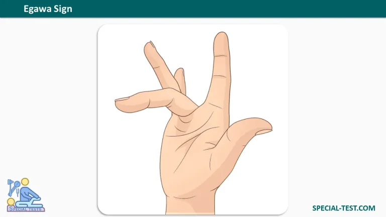
The Egawa sign is a specialized clinical examination maneuver used to assess the function of the intrinsic muscles of the hand—specifically the interossei—and to detect ulnar nerve palsy. Because the interosseous muscles are among the first to be affected in ulnar nerve compression, the Egawa sign can be invaluable for early diagnosis, often before more obvious deformities or sensory changes appear. This article explains the anatomy behind the test, how to perform it, how to interpret the result, and why it remains a trusted bedside tool for clinicians evaluating ulnar neuropathy.
What Is the Egawa Sign?
The Egawa sign (also called the Egawa test) is a provocative motor test designed to isolate and evaluate the interosseous muscles of the hand, all of which are innervated by the deep branch of the ulnar nerve. The test is performed by asking the patient to flex the middle finger at the metacarpophalangeal (MCP) joint and then actively deviate the finger radially and ulnarly—in other words, to abduct the finger in both directions while maintaining MCP flexion.
The scientific rationale is straightforward: the dorsal interossei are responsible for finger abduction, while the palmar interossei control finger adduction. When the MCP joint is held in flexion, the long extrinsic extensors cannot substitute for this side‑to‑side movement. Therefore, an inability to smoothly abduct and adduct the flexed middle finger points directly to interosseous weakness and, by extension, to ulnar nerve dysfunction.
How to Perform the Egawa Sign Test
The test is simple, quick, and requires no special equipment. A standardized technique improves reliability:
- Position the patient: The patient sits comfortably with the hand resting on a flat surface, palm down.
- Instruct the movement: Ask the patient to flex the middle finger at the MCP joint while keeping the interphalangeal (IP) joints extended.
- Test abduction and adduction: With the MCP joint held in flexion, instruct the patient to move the middle finger first toward the radial side (toward the thumb) and then toward the ulnar side (toward the little finger), alternating smoothly.
- Observe and compare: Watch for the ability to maintain stable MCP flexion while performing the side‑to‑side motion. Comparing both hands often reveals subtle asymmetry that may be missed on a single‑hand examination.
What Does a Positive Egawa Sign Indicate?
A negative (normal) Egawa sign means the patient can maintain MCP flexion and smoothly alternate radial and ulnar deviation without weakness or instability. This indicates intact interosseous muscle function.
A positive Egawa sign is present when the patient demonstrates any of the following:
- Inability to perform the alternating abduction/adduction movements.
- Weakness or a restricted range of deviation.
- Difficulty maintaining the required MCP flexion position.
- Tremor or instability during the attempted movement.
A positive result strongly suggests interosseous muscle compromise and is indicative of ulnar nerve palsy. Because the interossei are early targets in ulnar neuropathy, the Egawa sign can be positive even when grip strength and other routine measures remain relatively preserved.
Clinical Significance and Localization
The Egawa sign is more than a generic marker of ulnar neuropathy; it helps localize the lesion. The interosseous muscles are supplied by the deep motor branch of the ulnar nerve. A positive Egawa sign therefore points toward a lesion affecting this deep branch, whether at the elbow (cubital tunnel syndrome), at the wrist (Guyon’s canal syndrome), or along the motor branch in the palm. This distinction is clinically useful because sensory findings and other signs (such as Wartenberg’s sign) may differ depending on the level of compression.
The test is also valuable for serial monitoring. Clinicians can repeat the Egawa sign over time to track disease progression or recovery after conservative or surgical treatment. Its simplicity and low cost make it especially attractive in outpatient and primary‑care settings where advanced electrodiagnostic testing is not immediately available.
Differential Diagnosis
When the Egawa sign is positive, ulnar nerve palsy is the most likely explanation, but clinicians should also consider cubital tunnel syndrome, Guyon’s canal syndrome, and C8–T1 cervical radiculopathy, all of which can produce intrinsic hand weakness. Brachial plexopathy may also mimic the findings. Importantly, anterior interosseous nerve syndrome (a median nerve branch condition) typically affects different muscle groups and would not produce a positive Egawa sign, making the test useful for differentiation.
FAQ Section
Q1: What is the Egawa sign used for?
The Egawa sign is used to assess interosseous muscle function and to detect ulnar nerve palsy. It is particularly helpful for early diagnosis because the interossei are among the first muscles affected by ulnar nerve compression.
Q2: How is the Egawa sign performed?
The patient flexes the middle finger at the MCP joint while keeping the IP joints extended, then alternately deviates the finger radially and ulnarly. The examiner observes whether the patient can perform this movement smoothly and without weakness.
Q3: What does a positive Egawa sign mean?
A positive Egawa sign—difficulty or inability to abduct and adduct the flexed middle finger—indicates interosseous weakness and suggests ulnar nerve palsy.
Q4: Is the Egawa sign reliable for early ulnar neuropathy?
Yes. Because the interosseous muscles are early targets of ulnar nerve dysfunction, the Egawa sign can reveal weakness before more obvious signs such as clawing or marked grip loss appear.
References
- Orthofixar. Egawa Sign Test: Isolating High Vs. Low Ulnar Nerve Lesions. Available at: https://orthofixar.com/special-test/egawa-sign-test/
- Jennings JD, Jennings JF. Isolated Compression of the Ulnar Nerve Motor Branch: A Case Series With 3 Unique Etiologies. Ann Plast Surg. 2018;80(5):529-532. PMID: 29489540. Available at: https://pubmed.ncbi.nlm.nih.gov/29489540/
- Beutel BG, Worley C, Hosseinzadeh S. Intrinsic Hand Deformity. In: StatPearls [Internet]. Treasure Island (FL): StatPearls Publishing; 2025 Jan-. Available at: https://www.ncbi.nlm.nih.gov/books/NBK564355/
- Goldman SB, Brininger TL, Schrader JS, Koceja DM. A review of clinical tests and signs for the assessment of ulnar neuropathy. J Hand Ther. 2009;22(3):209-220. (Cited in Orthofixar)
- Signs and tests for ulnar nerve palsy. J Hand Surg. Table 1. Available at: https://www.jhandsurg.org/action/showFullTableHTML?isHtml=true&tableId=TBL1&pii=S1531-0914(03)50003-1