Biceps Reflex Test: How to Perform, C5–C6 Nerve Roots & Clinical Interpretation
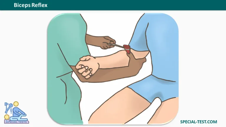
The Biceps Reflex Test, also known as the Biceps Tendon Reflex or C5–C6 Deep Tendon Reflex, is a neurological examination technique used to assess the integrity of the C5 and C6 spinal nerve roots, the musculocutaneous nerve, and the biceps brachii muscle.
This test is commonly performed during neurological and orthopedic examinations to evaluate the function of the cervical spinal cord and peripheral nervous system. It helps clinicians identify abnormalities associated with cervical radiculopathy, peripheral nerve injuries, and upper or lower motor neuron disorders.
The biceps reflex is one of the primary deep tendon reflexes (DTRs) examined in routine clinical practice, alongside the brachioradialis, triceps, patellar, and Achilles reflexes.
What Does the Biceps Reflex Test Assess?
The primary purpose of the biceps reflex test is to evaluate the neurological pathways responsible for the contraction of the biceps brachii muscle.
The test specifically examines:
- Nerve roots: C5 and C6, with C5 traditionally considered the predominant contribution.
- Peripheral nerve: Musculocutaneous nerve.
- Muscle tested: Biceps brachii.
- Reflex type: Deep tendon reflex (muscle stretch reflex).
- Expected response: Contraction of the biceps brachii muscle, resulting in slight elbow flexion.
The biceps reflex is mediated through a spinal reflex arc. When the biceps tendon is tapped, the sudden stretch activates muscle spindle receptors, generating sensory impulses that travel toward the spinal cord.
These signals activate motor neurons that stimulate the biceps brachii to contract.
An intact reflex indicates that the tested reflex pathway is functioning, although a normal result does not exclude every neurological disorder.
How to Perform the Biceps Reflex Test
The biceps tendon reflex test is performed using a neurological reflex hammer. Proper positioning, muscle relaxation, and correct percussion technique are essential for obtaining reliable results.
Patient Position
The patient is seated comfortably, with the upper limb relaxed and the elbow partially flexed.
The forearm should be supported, with the patient looking away from the tested arm to minimize anticipation and voluntary muscle contraction.
Examiner Position
The examiner stands beside the patient and supports the patient’s forearm with one hand.
The examiner places the thumb or finger gently over the biceps tendon in the antecubital fossa, just anterior to the elbow joint.
Testing Procedure
- Ask the patient to relax the arm completely.
- Support the forearm with the elbow positioned in partial flexion.
- Locate the biceps tendon in the anterior aspect of the elbow.
- Place the palmar surface of your thumb or finger lightly over the tendon.
- Using a reflex hammer, tap the examiner’s thumb or finger with a quick, controlled movement.
- Observe and palpate the biceps muscle for contraction.
- Repeat the examination on the opposite arm to compare the reflex responses.
Avoid applying excessive pressure over the tendon, because this may make the reflex more difficult to elicit.
Normal Biceps Reflex Response
A normal biceps reflex produces a brief contraction of the biceps brachii muscle, accompanied by slight flexion of the elbow.
A small amount of forearm supination may also occur because the biceps brachii contributes to both elbow flexion and forearm supination.
The strength of the reflex varies among individuals. Therefore, comparing the responses on both sides is an important component of the examination.
Biceps Reflex Grading Scale (0 to 4+)
Deep tendon reflexes are commonly graded using a 0 to 4+ scale, which describes the intensity of the response.
| Grade | Reflex Response | Clinical Interpretation |
|---|---|---|
| 0 | No detectable reflex response | Areflexia (absent reflex) |
| 1+ | Diminished or weak response, sometimes requiring reinforcement | Hyporeflexia |
| 2+ | Normal reflex response | Normal |
| 3+ | Brisk or exaggerated reflex response | May indicate hyperreflexia |
| 4+ | Very brisk reflex with repetitive contractions (clonus) | Markedly abnormal reflex response |
Important: A reflex graded 1+ or 3+ is not necessarily pathological. Interpretation depends on the patient’s baseline reflexes, symmetry, and associated neurological findings.
An absent or exaggerated reflex should be considered alongside muscle strength, sensation, muscle tone, and other examination results.
What Does an Abnormal Biceps Reflex Indicate?
Unlike some orthopedic special tests, the biceps reflex test does not have a single positive or negative result. Instead, clinicians evaluate whether the response is normal, diminished, absent, or exaggerated.
1. Hyporeflexia or Areflexia (0 to 1+)
A decreased or absent biceps reflex may indicate dysfunction involving the reflex arc.
Potential causes include:
- C5 or C6 cervical radiculopathy.
- Musculocutaneous nerve injury.
- Peripheral neuropathy.
- Lower motor neuron disorders.
For example, compression or irritation of a cervical nerve root can interfere with normal reflex transmission, potentially producing a reduced biceps tendon reflex on the affected side.
A unilateral decrease in reflex activity may be more clinically significant than symmetrical reductions, particularly when accompanied by muscle weakness or sensory changes.
2. Hyperreflexia (3+ to 4+)
An exaggerated biceps reflex may suggest dysfunction of descending motor pathways, including those involved in upper motor neuron control.
Possible associated conditions include:
- Cervical spinal cord compression (cervical myelopathy).
- Stroke affecting motor pathways.
- Multiple sclerosis.
- Other upper motor neuron disorders.
Hyperreflexia becomes more concerning when accompanied by spasticity, increased muscle tone, pathological reflexes, or neurological weakness.
However, a brisk reflex alone does not establish a neurological diagnosis.
Clinical Significance of the Biceps Reflex Test
The biceps reflex is particularly useful when evaluating patients with neck pain, arm weakness, numbness, or suspected cervical nerve root involvement.
Assessment of C5–C6 Radiculopathy
The biceps reflex helps assess the neurological integrity of the C5 and C6 spinal nerve roots.
A reduced reflex, particularly when accompanied by appropriate muscle weakness and sensory abnormalities, may support suspicion of cervical radiculopathy.
However, the biceps reflex cannot independently confirm or exclude nerve root compression.
A 2025 scoping review published in BMC Musculoskeletal Disorders found that reduced tendon reflexes can have relatively high specificity for cervical radiculopathy, although the overall evidence regarding neurological examination accuracy remains limited.
Differentiating Upper and Lower Motor Neuron Lesions
Deep tendon reflex testing contributes to identifying whether neurological dysfunction may involve the peripheral reflex arc or descending central motor pathways.
Decreased reflexes may occur with lower motor neuron dysfunction, whereas exaggerated reflexes may occur with upper motor neuron involvement.
The findings should always be interpreted within the complete neurological examination.
Common Mistakes During Biceps Reflex Testing
Several technical factors can affect the reliability of the examination:
- Excessive muscle tension: The patient should remain relaxed throughout the examination.
- Incorrect tendon localization: The examiner should identify the biceps tendon accurately before tapping.
- Excessive thumb pressure: Firm compression may interfere with the ability to elicit the reflex.
- Inadequate hammer technique: Percussion should be brisk, controlled, and directed over the tendon.
- Failure to compare sides: Bilateral examination helps identify clinically important asymmetry.
If the reflex is difficult to elicit, the examiner may repeat the test after adjusting the patient’s position or using an appropriate reinforcement maneuver.
Reflex grading is partly subjective. Research has demonstrated variability between examiners, making consistent testing technique and documentation important.
Key Clinical Takeaways
The biceps reflex test is a quick, inexpensive, and useful component of the neurological examination.
It primarily assesses the C5–C6 spinal nerve roots, musculocutaneous nerve, and biceps brachii muscle.
A normal response involves biceps contraction and slight elbow flexion. Diminished reflexes may suggest peripheral or lower motor neuron dysfunction, while exaggerated reflexes may suggest upper motor neuron involvement.
However, reflex findings should never be used in isolation to establish a diagnosis. Clinical interpretation requires correlation with the patient’s history, strength testing, sensory examination, and other neurological findings.
Frequently Asked Questions (FAQ)
1. What nerve roots are tested by the biceps reflex?
The biceps reflex primarily assesses the C5 and C6 spinal nerve roots, with C5 commonly regarded as the dominant contribution. The reflex travels through the musculocutaneous nerve to the biceps brachii muscle.
2. How do you perform the biceps reflex test?
Support the patient’s relaxed forearm, position your thumb or finger over the biceps tendon near the elbow, and gently strike your thumb or finger with a reflex hammer. Observe for biceps contraction and slight elbow flexion.
3. What is a normal biceps reflex?
A normal biceps reflex produces a brief contraction of the biceps brachii muscle and slight elbow flexion. It is typically graded 2+ on the deep tendon reflex grading scale.
4. What does an absent biceps reflex indicate?
An absent biceps reflex may suggest dysfunction of the C5–C6 nerve roots, musculocutaneous nerve, or other components of the peripheral reflex arc. However, further neurological examination is necessary to determine its significance.
5. What does an increased biceps reflex indicate?
An exaggerated biceps reflex may indicate upper motor neuron involvement, particularly when associated with increased muscle tone, weakness, or other pathological neurological signs.
6. Is the biceps reflex C5 or C6?
The biceps reflex involves both C5 and C6, although it is traditionally described as predominantly a C5 reflex. It is therefore commonly used during clinical assessment of both cervical nerve roots.
7. Can the biceps reflex test diagnose cervical radiculopathy?
No. A reduced or asymmetrical biceps reflex can support the clinical suspicion of cervical radiculopathy, but it cannot independently confirm the diagnosis. Muscle strength, sensation, clinical history, and additional investigations may be required.
8. What is the difference between the biceps and brachioradialis reflexes?
Both reflexes assess neurological pathways involving C5 and C6. However, the biceps reflex tests the biceps brachii through the musculocutaneous nerve, while the brachioradialis reflex tests the brachioradialis muscle through the radial nerve.
References
- Orthofixar – Biceps Reflex Test: Technique, Interpretation, and Clinical Significance
- Orthofixar – Deep Tendon Reflex Testing
- Walker HK. Deep Tendon Reflexes. Clinical Methods. PubMed PMID: 21250237
- Physiology, Deep Tendon Reflexes. PubMed PMID: 32965909
- Yousif MS, et al. Neurological Examination for Cervical Radiculopathy: A Scoping Review. 2025. PubMed PMID: 40188056
- Mayo and NINDS Scales for Assessment of Tendon Reflexes: Between Observer Agreement and Implications for Communication. PubMed PMID: 9489542