Tibialis Posterior Reflex Test: L5 Nerve Root Assessment, Technique & Interpretation
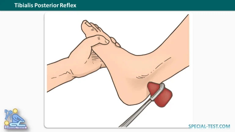
The Tibialis Posterior Reflex, also known as the posterior tibial tendon reflex, is a deep tendon reflex (DTR) used during neurological examinations to assess the integrity of the L5 nerve root and the associated peripheral nerve pathways. This reflex involves the tibialis posterior muscle, which is innervated by the tibial nerve.
The test is particularly useful as an additional clinical assessment in patients with suspected L5 radiculopathy, lumbar nerve root compression, or peripheral nerve dysfunction. By tapping the tibialis posterior tendon with a reflex hammer, the examiner evaluates the resulting involuntary muscle contraction and foot movement.
Although the tibialis posterior reflex can provide valuable neurological information, it is less consistently elicited than commonly tested reflexes such as the patellar and Achilles tendon reflexes. Therefore, findings must be interpreted alongside other components of the neurological examination.
What Is the Tibialis Posterior Reflex Used For?
The tibialis posterior reflex test primarily evaluates the neurological pathways associated with the L5 spinal nerve root. It may also provide information about the function of the tibial nerve and the tibial component of the sciatic nerve.
The main clinical applications include:
- L5 nerve root assessment: Helps identify possible neurological dysfunction associated with L5 radiculopathy.
- Tibial nerve evaluation: Assesses the integrity of the peripheral nerve supplying the tibialis posterior muscle.
- Lumbar radiculopathy examination: Provides supplementary information when investigating nerve root compression caused by lumbar spine disorders.
- Neurological screening: Helps identify asymmetrical, diminished, or exaggerated deep tendon reflex responses.
- Peripheral nerve assessment: Contributes to differentiating possible nerve root involvement from peripheral neuropathies when combined with motor and sensory testing.
Tibialis Posterior Reflex Innervation
| Anatomical Structure | Description |
|---|---|
| Reflex name | Tibialis Posterior Reflex |
| Muscle tested | Tibialis posterior |
| Peripheral nerve | Tibial nerve |
| Primary nerve root assessed | L5 |
| Additional segmental contributions | L4 and S1 may contribute |
| Tendon location | Posterior to the medial malleolus |
| Normal reflex response | Foot inversion, sometimes accompanied by slight plantar flexion |
The tibialis posterior muscle is located in the deep posterior compartment of the leg. Its primary functions include inversion of the foot, assistance with plantar flexion, and support of the medial longitudinal arch.
Because the muscle receives substantial L5 nerve root contributions, examination of its function can assist in the neurological assessment of patients with suspected lumbar radiculopathy.
How to Perform the Tibialis Posterior Reflex Test
Correct patient positioning, adequate muscle relaxation, and accurate tendon percussion are essential for obtaining a reliable reflex response.
1. Patient Position
Position the patient side-lying on the side being tested, with the test foot extending beyond the edge of the examination table.
The lower limb should remain relaxed, allowing the examiner to access the tibialis posterior tendon near the medial malleolus.
Alternatively, the test may be performed with the patient seated or prone, provided that the foot is adequately supported and the tendon can be accessed.
2. Examiner Position
The examiner stands or sits beside the patient, facing the medial aspect of the ankle.
The examiner may support the patient’s foot to maintain a comfortable position and minimize voluntary muscle activity.
3. Evaluative Procedure
- Identify the medial malleolus on the inner aspect of the ankle.
- Locate the tibialis posterior tendon immediately posterior to the medial malleolus.
- Position the foot in slight eversion, with gentle dorsiflexion if appropriate, to place the tendon under mild stretch.
- Ask the patient to relax the foot and ankle muscles completely.
- Using a neurological reflex hammer, deliver a quick, controlled tap over the tibialis posterior tendon, typically just posterior and proximal to the medial malleolus.
- Observe the foot for a brief involuntary inversion movement.
- Repeat the procedure on the opposite side to compare reflex intensity and symmetry.
Normal Response: A brief contraction of the tibialis posterior muscle produces inversion of the foot, occasionally accompanied by slight plantar flexion.
If the reflex is difficult to elicit, the examiner may repeat the test after repositioning the foot or using an appropriate reinforcement maneuver.
Tibialis Posterior Reflex Grading and Interpretation
The tibialis posterior reflex can be documented using the conventional 0 to 4+ deep tendon reflex grading scale.
| Grade | Reflex Response | Clinical Interpretation |
|---|---|---|
| 0 | No reflex elicited | Absent reflex |
| 1+ | Reduced or minimal response, sometimes elicited only with reinforcement | Hyporeflexia |
| 2+ | Normal reflex response | Normoreflexia |
| 3+ | Brisk or exaggerated response | Increased reflex activity; may be normal depending on context |
| 4+ | Markedly exaggerated response, potentially accompanied by clonus | Hyperreflexia |
A grade of 2+ generally indicates a normal reflex response. However, interpretation requires consideration of the patient’s baseline neurological function and comparison with the opposite limb.
A 1+ response is not necessarily pathological, especially when reflexes are symmetrically diminished. Similarly, a 3+ response may occur without neurological disease.
The standard grading system describes overall deep tendon reflex activity. Its application to this less consistently elicited reflex should be interpreted cautiously.
What Is a Positive Tibialis Posterior Reflex Test?
Unlike many orthopedic special tests, the tibialis posterior reflex does not have a single positive or negative result.
Instead, the examiner evaluates whether the reflex is present, absent, diminished, exaggerated, or asymmetrical.
Absent or Reduced Tibialis Posterior Reflex
A decreased or absent response may raise suspicion of dysfunction involving the reflex arc, including:
- L5 nerve root involvement.
- Tibial nerve injury or neuropathy.
- Sciatic nerve dysfunction.
- Other peripheral neurological disorders.
However, an absent tibialis posterior reflex alone does not establish a diagnosis of L5 radiculopathy.
The reflex can be difficult to elicit even in individuals without neurological disease. Therefore, unilateral loss or a marked difference between sides may be more informative than bilateral absence.
Exaggerated Tibialis Posterior Reflex
An unusually brisk reflex may suggest increased reflex excitability, particularly when accompanied by other upper motor neuron signs.
Marked hyperreflexia and clonus can indicate dysfunction of descending motor pathways, although these findings must be considered in the broader neurological examination.
Clinical Significance in L5 Radiculopathy
L5 radiculopathy occurs when the fifth lumbar spinal nerve root becomes irritated or compressed. It may be associated with lumbar disc herniation, degenerative spinal changes, or narrowing of the neural foramen.
Potential clinical findings include radiating leg pain, altered sensation, and weakness affecting muscles supplied by the L5 nerve root.
The tibialis posterior reflex may contribute to the neurological examination, but its diagnostic value is limited by variability in eliciting a response.
Research published in Surgical Neurology International emphasizes that the tibialis posterior reflex is not consistently present in healthy individuals and discusses the medial hamstring reflex as another assessment option for suspected L5 radiculopathy.
For a more comprehensive examination, clinicians should combine reflex testing with:
- Manual muscle testing of ankle inversion and great toe extension.
- Sensory examination of the lower limb and foot.
- Medial hamstring reflex assessment.
- Patellar and Achilles tendon reflex testing.
- Additional investigations, including electrodiagnostic studies or imaging, when clinically indicated.
Clinical Pearl: A unilateral reduction or absence of the tibialis posterior reflex, particularly when associated with L5-pattern muscle weakness or sensory changes, may support suspicion of L5 nerve root dysfunction. However, no isolated reflex finding should be used as definitive diagnostic evidence.
Frequently Asked Questions (FAQ)
1. Which nerve root is tested by the tibialis posterior reflex?
The tibialis posterior reflex primarily assesses the L5 nerve root, although additional segmental contributions may involve L4 and S1. The peripheral nerve responsible for innervating the tibialis posterior muscle is the tibial nerve.
2. What is the normal response to the tibialis posterior reflex?
The normal response is a brief involuntary contraction of the tibialis posterior muscle, resulting in inversion of the foot. Slight plantar flexion may also occur. The examiner should compare the response with the opposite limb.
3. Where should the reflex hammer strike during the test?
The examiner taps the tibialis posterior tendon near the medial malleolus, typically just posterior and slightly proximal to it. The tendon may also be accessed along its course behind the medial malleolus.
4. What does an absent tibialis posterior reflex indicate?
An absent response may be associated with L5 nerve root dysfunction, tibial nerve injury, or other abnormalities affecting the reflex pathway. However, this reflex is not consistently elicited in healthy people, so bilateral absence is not diagnostic by itself.
5. Is the tibialis posterior reflex useful for diagnosing L5 radiculopathy?
Yes, it can provide supplementary information during the examination of suspected L5 radiculopathy. However, it should be interpreted alongside muscle strength, sensory findings, other reflexes, and the patient’s clinical history.
6. What is the difference between the tibialis posterior reflex and the Achilles reflex?
The tibialis posterior reflex primarily assesses L5-related pathways and produces foot inversion. The Achilles tendon reflex predominantly assesses the S1 nerve root, with S2 contributions, and produces plantar flexion through contraction of the gastrocnemius and soleus muscles.
7. What does a 1+ tibialis posterior reflex mean?
A 1+ response indicates a diminished reflex. It may represent a normal variation or reduced reflex activity associated with neurological dysfunction. Reinforcement may be required to elicit a detectable response.
8. Can a normal tibialis posterior reflex rule out L5 radiculopathy?
No. A normal reflex does not exclude L5 radiculopathy. Reflex testing is only one component of the neurological examination, and patients with nerve root compression may have preserved reflex responses.
Conclusion
The Tibialis Posterior Reflex Test is an additional neurological assessment used to investigate L5 nerve root function and the integrity of tibial nerve pathways. The test involves tapping the tibialis posterior tendon near the medial malleolus and observing for involuntary foot inversion.
Proper positioning, muscle relaxation, bilateral comparison, and accurate reflex grading help improve clinical interpretation.
Because the reflex is not consistently elicited in all healthy individuals, abnormal findings should be evaluated alongside muscle strength, sensation, other deep tendon reflexes, and the overall clinical presentation.
References
- Orthofixar. Tibialis Posterior Reflex Test: How to Assess L5 Nerve Root Function.
- Orthofixar. Deep Tendon Reflex Testing: Grading, Technique & Nerve Root Guide.
- Deep Tendon Reflex: The Tools and Techniques. What Surgical Neurology Residents Should Know. Published in Surgical Neurology International, 2021. NCBI PubMed Central.
- Esene IN, et al. (2012). Diagnostic performance of the medial hamstring reflex in L5 radiculopathy. Surgical Neurology International, 3:104. NCBI PubMed Central.
- Zimmerman B, Hubbard JB. Deep Tendon Reflexes. StatPearls. NCBI Bookshelf.
- Deep Tendon Reflexes. Clinical Methods: The History, Physical, and Laboratory Examinations. NCBI Bookshelf.
- Corcoran NM, Varacallo MA. Anatomy, Bony Pelvis and Lower Limb: Tibialis Posterior Muscle. StatPearls. NCBI Bookshelf.
- Röder H, Lang G. (1977). Measurement of muscle reflex time in radicular disorders of the lumbosacral region. PubMed PMID: 202981.
- Heffernan LP. (1979). Electromyographic value of the tibialis posterior muscle. Archives of Physical Medicine and Rehabilitation. PubMed PMID: 224837.