Biceps Femoris Reflex Test: How to Perform, Nerve Roots & Interpretation
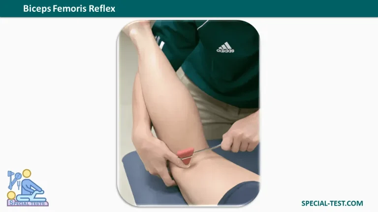
What Is the Biceps Femoris Reflex?
The Biceps Femoris Reflex, also known as the Lateral Hamstring Reflex or External Hamstring Reflex, is a deep tendon reflex used during neurological and musculoskeletal examinations to evaluate the integrity of the lower-limb reflex pathways.
The test involves tapping the biceps femoris tendon near the posterolateral aspect of the knee to produce an involuntary contraction of the hamstring muscle. The expected response is a brief contraction of the biceps femoris, which may produce visible knee flexion.
The biceps femoris reflex is particularly useful when assessing suspected S1 nerve root dysfunction, lumbar radiculopathy, sciatic nerve abnormalities, and other neurological conditions affecting the lower extremities.
Although the biceps femoris muscle receives contributions from several lumbosacral nerve roots, the lateral hamstring reflex is primarily associated with the S1 spinal segment.
Biceps Femoris Reflex: Nerve Root and Innervation
The biceps femoris is one of the major hamstring muscles located in the posterior compartment of the thigh. It consists of two heads that receive innervation from different divisions of the sciatic nerve.
- Long head: Innervated by the tibial division of the sciatic nerve.
- Short head: Innervated by the common fibular (common peroneal) division of the sciatic nerve.
- Spinal nerve roots: The muscle receives contributions primarily from L5, S1, and S2.
- Primary reflex level: S1.
- Main reflex response: Hamstring contraction with knee flexion.
The biceps femoris reflex is commonly used as an additional assessment of the S1 nerve root, particularly when findings from the Achilles tendon reflex are unclear.
The anatomical innervation of the entire muscle should not be confused with the dominant spinal segment tested by the lateral hamstring reflex.
How to Perform the Biceps Femoris Reflex Test
The Biceps Femoris Reflex Test is performed using a neurological reflex hammer. Accurate patient positioning, appropriate tendon localization, and muscle relaxation are essential for obtaining a reliable response.
1. Patient Position
Position the patient in a prone position, lying face down on an examination table.
- Keep the patient comfortable and relaxed.
- Flex the knee to approximately 20 degrees.
- Ensure the hamstring muscles are not actively contracting.
- Support the limb as necessary to allow an unrestricted reflex response.
Alternatively, the examination may be performed with the patient seated, recumbent, or side-lying with the knee moderately flexed.
2. Examiner Position
The examiner stands beside the patient’s lower limb, facing the posterolateral aspect of the knee.
The biceps femoris tendon is located near the lateral side of the popliteal region, immediately proximal to its insertion around the head of the fibula.
Place the thumb or fingers over the palpable tendon just above the knee joint line.
3. Evaluative Procedure
- Identify and palpate the biceps femoris tendon on the posterolateral side of the knee.
- Position the thumb firmly but comfortably over the tendon.
- Use a reflex hammer to deliver a quick, controlled tap to the examiner’s thumb.
- Observe or palpate the biceps femoris muscle for an involuntary contraction.
- Look for a brief knee-flexion response.
- Repeat the procedure on the opposite leg to compare reflex symmetry.
An alternative method involves tapping near the head of the fibula to elicit the lateral hamstring reflex.
4. Expected Normal Response
A normal biceps femoris reflex produces a brief, involuntary contraction of the hamstring muscle, sometimes accompanied by visible knee flexion.
The intensity of the response may vary among healthy individuals. Therefore, comparing the right and left sides is an important part of the assessment.
Biceps Femoris Reflex Grading and Interpretation
The biceps femoris reflex is evaluated using the standard deep tendon reflex grading system, which typically ranges from 0 to 4+.
| Grade | Reflex response | Clinical interpretation |
|---|---|---|
| 0 | No reflex elicited | Absent reflex (areflexia) |
| 1+ | Reduced or minimal response, sometimes requiring reinforcement | Hyporeflexia |
| 2+ | Normal reflex response | Normoreflexia |
| 3+ | Brisk or exaggerated response | Increased reflex activity; may be normal in some patients |
| 4+ | Markedly exaggerated response, potentially accompanied by clonus | Significant hyperreflexia; clonus is an abnormal finding |
A grade of 2+ is generally considered normal. However, isolated 1+ or 3+ responses do not necessarily indicate neurological disease.
Reflex grading should always be interpreted in relation to the patient’s symptoms, muscle strength, sensation, and other neurological findings.
What Is a Positive Biceps Femoris Reflex Test?
Unlike orthopedic special tests that produce a simple positive or negative result, deep tendon reflexes are assessed according to their intensity and symmetry.
An abnormal biceps femoris reflex may include an absent, diminished, unusually brisk, or asymmetric response.
Absent or Diminished Reflex
A reduced or absent biceps femoris reflex may suggest an abnormality involving the reflex arc, including:
- S1 nerve root dysfunction or radiculopathy.
- Peripheral nerve injury affecting the sciatic nerve.
- Lower motor neuron dysfunction.
- Peripheral neuropathy involving the relevant reflex pathway.
A unilateral reduction is generally more clinically significant than a symmetrical reduction when accompanied by corresponding weakness, sensory changes, or radiating leg pain.
Exaggerated Reflex
An unusually brisk response may indicate increased reflex excitability.
When accompanied by other neurological abnormalities, hyperreflexia may raise concern for upper motor neuron dysfunction.
However, a brisk reflex alone is insufficient to establish a neurological diagnosis.
Clinical Significance of the Biceps Femoris Reflex
The biceps femoris reflex can provide additional information during the evaluation of patients with lower back pain, sciatica, lower-limb weakness, or suspected lumbosacral radiculopathy.
Assessment of S1 Radiculopathy
The lateral hamstring reflex is primarily associated with S1 nerve root function.
In patients with suspected S1 radiculopathy, the clinician may assess both the biceps femoris and Achilles tendon reflexes.
Reduced reflex responses, particularly when associated with appropriate neurological symptoms, may support the suspicion of nerve root involvement.
Differentiating Radiculopathy From Peripheral Neuropathy
The lateral hamstring reflex may help distinguish certain patterns of peripheral neuropathy from nerve root dysfunction.
For example, a patient with a diminished Achilles tendon reflex but a preserved lateral hamstring reflex may have a different pattern of nerve involvement than a patient with both reflexes reduced.
Nevertheless, this distinction is not definitive and must be evaluated alongside the complete neurological examination.
Complementing Other Neurological Tests
The biceps femoris reflex may be assessed alongside:
- Patellar tendon reflex.
- Achilles tendon reflex.
- Lower-limb muscle strength testing.
- Dermatomal sensory examination.
- Straight leg raise testing when appropriate.
- Electrodiagnostic investigations when clinically indicated.
Combining these findings provides a more complete assessment of the lumbosacral nervous system.
Important Considerations During Reflex Testing
Several factors may influence the reliability of the examination.
Muscle relaxation: Voluntary muscle contraction may interfere with the reflex response. The patient should remain relaxed throughout the procedure.
Accurate tendon localization: The examiner must correctly identify the biceps femoris tendon near the lateral aspect of the knee.
Consistent technique: Use comparable tapping force and limb positioning when examining both legs.
Reinforcement: If the reflex is difficult to elicit, an appropriate reinforcement maneuver may be used. Any response obtained with reinforcement should be documented.
Bilateral comparison: Differences between the right and left sides may be more informative than the absolute reflex grade.
The biceps femoris reflex should never be interpreted in isolation when investigating neurological disease.
Frequently Asked Questions (FAQ)
1. What nerve root does the biceps femoris reflex test?
The biceps femoris reflex primarily evaluates the S1 nerve root, although the biceps femoris muscle receives additional segmental innervation, including L5 and S2. The lateral hamstring reflex is therefore most commonly associated with S1 rather than serving as an isolated S2 reflex test.
2. How do you test the biceps femoris reflex?
Position the patient prone with the knee slightly flexed. Palpate the biceps femoris tendon near the posterolateral knee, place your thumb over it, and tap your thumb using a reflex hammer. Observe or feel for hamstring contraction and knee flexion.
3. What is the normal biceps femoris reflex response?
The normal response is a brief, involuntary contraction of the biceps femoris muscle, which may produce slight knee flexion. A reflex grade of 2+ is conventionally considered normal.
4. What does an absent biceps femoris reflex indicate?
An absent reflex may suggest dysfunction involving the S1 nerve root, sciatic nerve, or another component of the reflex arc. However, technical factors and individual variation must be considered before interpreting the finding as pathological.
5. Is the biceps femoris reflex the same as the Achilles reflex?
No. Although both reflexes can assess S1-related neurological function, they involve different muscles and tendons.
The biceps femoris reflex is elicited from the lateral hamstring tendon, while the Achilles reflex is elicited from the Achilles tendon and produces plantar flexion of the ankle.
6. Can the biceps femoris reflex detect lumbar radiculopathy?
The reflex may provide supporting evidence for suspected lumbosacral radiculopathy, particularly S1 involvement. However, the test alone cannot confirm or exclude radiculopathy. Diagnosis requires correlation with the patient’s history, neurological examination, and additional investigations when indicated.
7. What is the difference between the lateral and medial hamstring reflexes?
The lateral hamstring reflex primarily involves the biceps femoris tendon and is commonly associated with S1.
The medial hamstring reflex evaluates a different hamstring reflex pathway and has been studied as a clinical indicator of L5 radiculopathy.
Distinguishing these reflexes is important because they may provide complementary information about lumbosacral nerve root function.
8. What does a 4+ biceps femoris reflex mean?
A 4+ response indicates markedly increased reflex activity. In commonly used clinical grading systems, it may be accompanied by clonus, which refers to repetitive involuntary muscular contractions.
When present, clonus warrants further neurological assessment.
Conclusion
The Biceps Femoris Reflex Test is a useful component of the lower-limb neurological examination. It primarily evaluates the S1 reflex pathway and can provide additional clinical information when assessing lumbar radiculopathy, sciatic nerve dysfunction, or abnormal deep tendon reflexes.
Correct positioning, precise tendon palpation, appropriate reflex hammer technique, and bilateral comparison are essential for reliable assessment.
Although abnormal reflex findings may suggest neurological dysfunction, results must always be interpreted alongside muscle strength, sensation, other reflexes, and the patient’s clinical presentation.
References
- Orthofixar. Biceps Femoris Reflex: Clinical Testing and S1 Nerve Root Evaluation. https://orthofixar.com/special-test/biceps-femoris-reflex/
- Orthofixar. Deep Tendon Reflex Testing: Grading, Technique & Nerve Root Guide. https://orthofixar.com/special-test/deep-tendon-reflex-testing/
- Deep Tendon Reflex: The Tools and Techniques. What Surgical Neurology Residents Should Know. PubMed Central (PMC). https://pmc.ncbi.nlm.nih.gov/articles/PMC8075597/
- Zimmerman B, Hubbard JB. Deep Tendon Reflexes. StatPearls. NCBI Bookshelf. https://www.ncbi.nlm.nih.gov/books/NBK531502/
- Anatomy, Bony Pelvis and Lower Limb: Femoral Muscles. StatPearls. NCBI Bookshelf. https://www.ncbi.nlm.nih.gov/books/NBK500008/
- Potential Advantages of the H-Reflex of the Biceps Femoris-Long Head in Documenting S1 Radiculopathy. PubMed. https://pubmed.ncbi.nlm.nih.gov/24492445/
- Diagnostic Performance of the Medial Hamstring Reflex in L5 Radiculopathy. PubMed. https://pubmed.ncbi.nlm.nih.gov/23087820/