Wright’s Test (Hyperabduction Test): Purpose, Procedure, Positive Result, and Diagnostic Accuracy
What Is Wright’s Test?
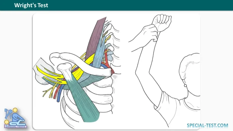
Wright’s Test, also known as the Hyperabduction Test, is a clinical orthopedic examination used to assess possible thoracic outlet syndrome (TOS). The test evaluates whether positioning the upper extremity in shoulder abduction produces neurovascular compression, potentially affecting the brachial plexus or blood vessels supplying the arm.
Thoracic outlet syndrome occurs when nerves or blood vessels become compressed as they pass through anatomical spaces between the neck, clavicle, first rib, and surrounding muscles.
Wright’s Test primarily investigates compression associated with the retropectoralis minor (subcoracoid) space, where the pectoralis minor muscle may contribute to compression of neurovascular structures during overhead arm movements.
Healthcare professionals use this provocative examination as part of a comprehensive assessment of patients experiencing upper extremity pain, numbness, tingling, weakness, or symptoms aggravated by raising the arm.
What Is Wright’s Test Used For?
The primary purpose of Wright’s Test is to assess patients with suspected thoracic outlet syndrome, particularly those whose symptoms become worse during shoulder elevation or overhead activities.
The test may help identify positional compression involving the brachial plexus or vascular structures.
Thoracic outlet syndrome is generally classified into three main types:
- Neurogenic thoracic outlet syndrome: Involves compression or irritation of the brachial plexus, potentially producing pain, paresthesia, weakness, and sensory disturbances.
- Venous thoracic outlet syndrome: Involves compression of the subclavian vein and may cause upper extremity swelling, discoloration, and venous congestion.
- Arterial thoracic outlet syndrome: Involves compression or damage to the subclavian artery and may produce ischemic symptoms, including hand pain, coolness, or color changes.
Wright’s Test can help provoke symptoms associated with positional neurovascular compression. However, it cannot reliably distinguish these three conditions or establish the diagnosis independently.
How to Perform Wright’s Test
The Wright’s Test procedure should be performed by a trained healthcare professional during an appropriate physical examination.
Step-by-Step Procedure
- Patient position: Ask the patient to sit or stand comfortably with their shoulders relaxed and arms resting alongside the body.
- Locate the radial pulse: The examiner palpates the radial artery at the wrist of the extremity being assessed to establish the baseline pulse.
- Abduct the shoulder: While monitoring the radial pulse, the examiner gently and passively abducts the patient’s arm. In the traditional hyperabduction maneuver, the arm is progressively elevated toward approximately 180 degrees, as tolerated.
- Assess the response: Observe whether the radial pulse weakens or disappears during shoulder abduction. Ask the patient whether the maneuver reproduces their usual symptoms, such as numbness, tingling, pain, or weakness.
- Compare both sides: Repeat the examination on the opposite upper extremity and document any differences in pulse response or symptom reproduction.
Modified Wright’s Test
A commonly described variation involves passive shoulder abduction and external rotation, often with the elbow flexed, while the patient turns their head away from the tested extremity.
During this modified maneuver, the examiner continues monitoring the radial pulse and asks about neurological symptoms.
This variation is sometimes described as the Allen maneuver and should be distinguished from the traditional Wright hyperabduction procedure.
The examiner should avoid forcing the shoulder into a painful position or unnecessarily prolonging the maneuver.
What Is a Positive Wright’s Test?
A positive Wright’s Test traditionally occurs when the radial pulse becomes noticeably weaker or disappears during shoulder hyperabduction.
Other findings that may be clinically relevant include reproduction of the patient’s familiar symptoms, such as:
- Numbness or tingling in the arm or hand.
- Upper extremity pain or discomfort.
- A sensation of the hand falling asleep.
- Weakness or heaviness during arm elevation.
- Changes in hand temperature or coloration.
These responses may suggest positional neurovascular compression.
However, a diminished or absent radial pulse does not necessarily confirm thoracic outlet syndrome. Pulse changes during provocative arm positioning have also been documented in healthy individuals.
Reproduction of the patient’s typical neurological symptoms may provide additional clinical information, particularly when interpreted alongside the history and other examination findings.
Wright’s Test Sensitivity and Specificity
The diagnostic accuracy of Wright’s Test remains uncertain.
Although sensitivity and specificity values of 32% and 96%, respectively, are sometimes reported in educational summaries, these particular estimates could not be verified as established values for Wright’s Test using the medical references reviewed here.
Orthofixar states that sufficiently established diagnostic accuracy data are unavailable for assigning reliable sensitivity and specificity values to the maneuver.
Furthermore, research demonstrates that positive vascular responses may occur in individuals without thoracic outlet syndrome.
In a study published in the Journal of Shoulder and Elbow Surgery, Rayan and Jensen examined 200 upper extremities from 100 volunteers. A vascular response during the hyperabduction maneuver occurred in 57% of extremities, while a neurological response occurred in 16.5%.
These results demonstrate an important limitation: radial pulse changes during hyperabduction are not specific to symptomatic thoracic outlet syndrome.
A systematic review by Dessureault-Dober and colleagues, published in the Journal of Manipulative and Physiological Therapeutics, also concluded that evidence supporting the diagnostic validity of clinical tests for thoracic outlet syndrome remains limited.
Consequently, Wright’s Test should be regarded as a supportive provocative examination rather than a definitive diagnostic test.
Clinical Significance of Wright’s Test
Wright’s Test is clinically useful because overhead arm positioning can reproduce symptoms experienced by individuals with suspected thoracic outlet syndrome.
The maneuver may provide information about symptom aggravation during shoulder elevation and possible compression in the region beneath the pectoralis minor muscle.
Nevertheless, clinicians must interpret the results carefully because several disorders can cause similar symptoms, including:
- Cervical radiculopathy.
- Peripheral nerve entrapment.
- Brachial plexus disorders.
- Shoulder musculoskeletal conditions.
- Other upper extremity vascular abnormalities.
A comprehensive diagnostic assessment includes medical history, neurological examination, vascular examination, and additional investigations when indicated.
Depending on the clinical presentation, appropriate investigations may include duplex ultrasonography, cervical or chest radiographs, electrodiagnostic studies, CT angiography, or magnetic resonance imaging.
Patients with acute arm swelling, pronounced color changes, severe pain, or signs of impaired blood circulation require prompt medical evaluation rather than reliance on provocative testing alone.
Key Clinical Points
Wright’s Test is a practical bedside maneuver for assessing suspected thoracic outlet syndrome, particularly when symptoms are associated with shoulder abduction and overhead activities.
The examination involves passive arm hyperabduction while monitoring the radial pulse and the patient’s symptoms. A modified version may incorporate external rotation and contralateral neck rotation.
A positive finding traditionally includes radial pulse reduction or disappearance, although reproduction of characteristic neurological symptoms can also be clinically relevant.
Because false-positive responses occur in healthy individuals and diagnostic accuracy remains uncertain, the test should always be interpreted as part of a broader clinical assessment.
Frequently Asked Questions (FAQ)
1. What does Wright’s Test assess?
Wright’s Test assesses possible positional neurovascular compression associated with thoracic outlet syndrome. It evaluates changes in the radial pulse and reproduction of symptoms during shoulder hyperabduction.
2. How do you perform Wright’s Test for thoracic outlet syndrome?
The examiner palpates the patient’s radial pulse while passively abducting the affected arm toward an overhead position. Pulse changes and neurological symptoms are monitored. A modified version may include external shoulder rotation and turning the neck away from the tested side.
3. What indicates a positive Wright’s Test?
A positive Wright’s Test traditionally involves a noticeable reduction or disappearance of the radial pulse during shoulder hyperabduction. Reproduction of familiar neurological symptoms, including numbness, tingling, or pain, is also clinically relevant.
4. Is Wright’s Test reliable for diagnosing thoracic outlet syndrome?
Wright’s Test has limited diagnostic reliability when used alone. Studies have demonstrated positive responses in asymptomatic individuals, and current evidence does not establish consistently reliable sensitivity and specificity values.
5. What is the difference between Wright’s Test and Adson’s Test?
Wright’s Test primarily uses shoulder hyperabduction to provoke neurovascular symptoms. Adson’s Test emphasizes neck positioning, deep inspiration, and radial pulse monitoring. Both are provocative examinations used in the assessment of suspected thoracic outlet syndrome, but neither should independently establish the diagnosis.
6. Can Wright’s Test detect neurogenic thoracic outlet syndrome?
Wright’s Test may reproduce symptoms associated with neurogenic thoracic outlet syndrome, such as paresthesia, discomfort, or weakness. However, it cannot reliably confirm neurogenic thoracic outlet syndrome or distinguish it from other causes of upper extremity neurological symptoms.
7. Does losing the radial pulse during Wright’s Test always mean thoracic outlet syndrome?
No. A diminished or absent radial pulse can occur during shoulder hyperabduction in otherwise healthy individuals. Therefore, pulse disappearance alone is insufficient to diagnose thoracic outlet syndrome.
8. Why is Wright’s Test also called the Hyperabduction Test?
The name refers to the primary examination movement: passive elevation of the arm into substantial shoulder abduction. This position can narrow the space around neurovascular structures beneath the pectoralis minor muscle and may provoke symptoms associated with thoracic outlet compression.
References
- Orthofixar. Wright Test: Hyperabduction Test for Thoracic Outlet Syndrome. Orthofixar – Wright Test.
- Rayan GM, Jensen C. Thoracic outlet syndrome: provocative examination maneuvers in a typical population. Journal of Shoulder and Elbow Surgery. 1995;4(2):113–117. PubMed PMID: 7600161.
- Dessureault-Dober I, Bronchti G, Bussières A. Diagnostic Accuracy of Clinical Tests for Neurogenic and Vascular Thoracic Outlet Syndrome: A Systematic Review. Journal of Manipulative and Physiological Therapeutics. 2018;41(9):789–799. PubMed PMID: 30871714.
- The Diagnostic Accuracy of Clinical Diagnostic Tests for Thoracic Outlet Syndrome. PubMed PMID: 27632823.
- Rayan GM. Thoracic outlet syndrome. Journal of Shoulder and Elbow Surgery. 1998;7(4):440–451. PubMed PMID: 9752662.