Deltoid Extension Lag Sign: Test for Axillary Nerve Injury
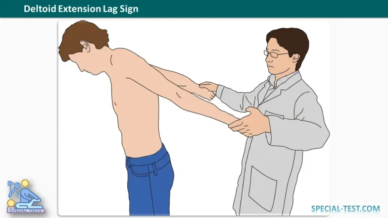
The deltoid extension lag sign is a clinical examination test used to assess posterior deltoid function and help identify axillary nerve palsy. It is particularly relevant when evaluating patients following traumatic shoulder injuries, including anterior glenohumeral dislocation.
Originally described by Hertel and colleagues in 1998, the test evaluates the patient’s ability to actively maintain shoulder extension after the examiner positions the arm. An inability to maintain the position may indicate posterior deltoid weakness associated with axillary nerve dysfunction. [1]
What Is the Deltoid Extension Lag Sign Used For?
The deltoid extension lag sign is used to assess posterior deltoid function and detect possible axillary nerve injury.
The axillary nerve arises from the posterior cord of the brachial plexus, primarily carrying fibers from the C5 and C6 nerve roots. It provides motor innervation to the deltoid and teres minor muscles and sensory innervation to the lateral shoulder.
Axillary nerve injuries may occur following:
- Anterior glenohumeral dislocation.
- Proximal humerus fractures.
- Direct trauma to the shoulder.
- Traction or compression injuries affecting the nerve.
The deltoid extension lag sign can also help clinicians monitor changes in deltoid function during recovery from an axillary nerve injury. However, it should be interpreted alongside the patient’s history and a comprehensive shoulder and neurological examination. [1, 2]
How to Perform the Deltoid Extension Lag Test
Patient position: The patient sits or stands comfortably, with the trunk kept upright.
Examination technique:
- Examine the unaffected side first. This establishes a baseline for comparison.
- Position the arm. The examiner passively moves the patient’s arm into near-maximal shoulder extension, keeping the elbow extended.
- Ask the patient to maintain the position. The patient actively holds the arm in the examiner-established position.
- Observe for extension lag. Note whether the patient can maintain the position or whether the arm drops from the target position.
- Repeat on the affected side. Compare the degree of extension lag and the patient’s ability to maintain the position between both shoulders.
Avoid compensatory trunk movement, particularly forward flexion, which may affect the validity of the examination.
The examiner should document any observed lag and consider pain, restricted shoulder mobility, and other injuries that may interfere with the patient’s performance. [1]
What Is a Positive Deltoid Extension Lag Sign?
A positive deltoid extension lag sign occurs when the patient cannot actively maintain the examiner-established shoulder extension position and the arm drops from that position.
A greater extension lag on the symptomatic side compared with the unaffected side suggests impaired posterior deltoid function and raises suspicion of axillary nerve palsy.
The original study by Hertel and colleagues reported that the magnitude of the arm’s angular drop correlated with deltoid functional status in a small series of patients with traumatic axillary nerve palsy following anterior shoulder dislocation. The authors also described the test’s potential for monitoring recovery over time. [1]
Clinical Interpretation
A positive test may indicate:
- Posterior deltoid weakness or dysfunction.
- Possible axillary nerve injury.
- Persistent impairment of deltoid function during follow-up after shoulder trauma.
Important: A positive test does not independently confirm an axillary nerve lesion. Pain, restricted movement, associated muscle or tendon injuries, and other neurological disorders may influence the findings.
Clinical Significance of the Deltoid Extension Lag Sign
Axillary nerve injury is an important complication of anterior shoulder dislocation and proximal humerus fracture. Because the nerve supplies the deltoid muscle, injury may result in weakness of shoulder abduction and impaired shoulder function.
Clinical assessment should include deltoid contraction, shoulder movement, and sensation over the lateral shoulder. Preserved sensation does not completely exclude axillary motor nerve injury.
Depending on the clinical presentation, further assessment may include radiographs to evaluate associated fractures, electrodiagnostic studies such as electromyography (EMG) and nerve conduction studies, or additional imaging. The choice and timing of investigations depend on the injury and clinical findings. [2, 3]
Following an acute shoulder dislocation, a documented neurovascular examination is essential. The deltoid extension lag sign is one component of the examination and should not delay urgent assessment or appropriate management of a suspected dislocation or associated fracture.
Frequently Asked Questions (FAQ)
1. What does the deltoid extension lag sign test?
The test assesses posterior deltoid function and helps identify weakness that may be associated with axillary nerve injury. It evaluates the patient’s ability to maintain an examiner-established shoulder extension position.
2. What nerve is affected when the deltoid extension lag sign is positive?
The test primarily raises suspicion of axillary nerve dysfunction, which can impair deltoid muscle function. However, a positive result is not specific enough to establish the diagnosis without further clinical assessment.
3. Why is the deltoid extension lag sign important after anterior shoulder dislocation?
The axillary nerve can be stretched or injured during anterior glenohumeral dislocation. Assessing deltoid function can help clinicians identify a possible motor deficit and monitor functional recovery.
4. How is a positive deltoid extension lag sign identified?
The patient is unable to maintain the shoulder extension position established by the examiner, and the arm drops. A greater lag on the affected side than on the unaffected side supports the presence of impaired deltoid function.
5. Does a positive deltoid extension lag sign confirm axillary nerve palsy?
No. The test supports clinical suspicion but does not independently confirm the diagnosis. A comprehensive examination should assess deltoid strength, shoulder movement, sensation, and possible associated injuries. EMG and nerve conduction studies may be indicated when further diagnostic clarification is needed.
6. Can the deltoid extension lag sign be used to monitor recovery?
Yes. Repeating the examination and documenting the degree of lag may help track changes in deltoid function over time. The original study described the test as useful for assessing functional recovery, although its findings were based on a small patient series.
7. What other clinical findings suggest axillary nerve injury?
Common findings include deltoid weakness, reduced shoulder abduction strength, and altered sensation over the lateral shoulder. These findings should be interpreted in the context of the injury and the overall neurological examination.
8. What investigations can help confirm axillary nerve injury?
Depending on the clinical context, investigations may include EMG, nerve conduction studies, and imaging. Radiographs are important when a fracture or traumatic shoulder dislocation is suspected. Electrodiagnostic studies can help assess nerve dysfunction and severity, but their timing after injury matters. [2, 3]
References
- Hertel R, Lambert SM, Ballmer FT. The deltoid extension lag sign for diagnosis and grading of axillary nerve palsy. Journal of Shoulder and Elbow Surgery. 1998;7(2):97–99. Read the original study on PubMed.
- Axillary Nerve Injury. StatPearls. National Center for Biotechnology Information (NCBI Bookshelf). Read the clinical review.
- Axillary nerve injury associated with glenohumeral dislocation: A review and algorithm for management. Read the peer-reviewed review on PubMed Central.