Withdrawal Reflex: Definition, How to Perform the Test, and Clinical Significance
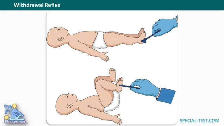
The withdrawal reflex, also known as the flexion withdrawal reflex, is an involuntary neurological response that protects the body from potentially harmful stimuli. In newborns and young infants, this reflex can be assessed during a neurological examination to evaluate the sensory and motor responses of the lower limbs and provide information about spinal cord function.
The withdrawal reflex occurs when a noxious stimulus, such as a painful stimulus applied to the sole of the foot, triggers a rapid flexion response that moves the affected limb away from the stimulus. The response involves sensory nerves, spinal cord circuits, and motor nerves that activate the muscles responsible for withdrawing the limb.
Clinical assessment of this reflex is particularly relevant during the neurological examination of infants with suspected spinal cord abnormalities, congenital neurological conditions, or disorders affecting muscle tone. However, the withdrawal reflex should be interpreted alongside other neurological findings rather than used as an isolated diagnostic test.
What Is the Withdrawal Reflex Used For?
The withdrawal reflex is assessed primarily as part of the neurological examination of newborns and infants. It helps clinicians evaluate the integrity of the sensory pathways, spinal reflex circuits, and motor pathways involved in withdrawing the lower limb from a harmful stimulus.
The main clinical purposes of assessing the withdrawal reflex include:
- Evaluating spinal cord function: The reflex provides information about the neural pathways responsible for the protective flexion response.
- Assessing sensory-motor responses: It demonstrates whether a stimulus applied to the foot produces an appropriate motor response.
- Identifying possible neurological abnormalities: A weak, absent, or atypical response may warrant further evaluation for neurological or spinal cord disorders.
- Supporting the assessment of congenital conditions: The reflex may be considered when examining infants with myelomeningocele or other spinal cord abnormalities.
- Contributing to developmental neurological assessment: The response can be interpreted alongside muscle tone, spontaneous movements, tendon reflexes, and other age-appropriate neurological findings.
Research on cutaneous withdrawal reflexes in human neonates demonstrates that these responses change with maturation and are influenced by gestational age, stimulus characteristics, and the infant’s neurological state. Therefore, the findings must be interpreted in the context of the infant’s age and overall clinical condition.
When Does the Withdrawal Reflex Appear and Disappear?
The withdrawal response is present at birth and can be elicited in newborns, including premature infants. The supplied clinical description places its expected disappearance between 1 and 2 months of age.
However, the flexion withdrawal reflex is a spinal protective response rather than a simple primitive reflex that necessarily disappears at a single, fixed age. Research has demonstrated that withdrawal responses continue to develop after birth, with changes in sensitivity and spinal cord excitability during infancy. Consequently, the response may remain demonstrable beyond the first two months, depending on how it is elicited and measured.
A clinician should therefore avoid diagnosing a neurological disorder solely because a withdrawal response remains present after 2 months of age. The infant’s developmental stage, the examination technique, and other neurological findings are essential to interpretation.
How to Perform the Withdrawal Reflex Test
The withdrawal reflex is assessed during a structured neurological examination. The examiner should use an appropriate clinical technique and avoid unnecessary painful stimulation.
Patient Position
- Place the infant in the supine position, lying comfortably on the back.
- Position the lower limbs near the midline in their natural resting alignment.
- Allow the legs to remain in a relaxed, natural position, without forcing the hips or knees into extension.
- Observe the infant’s spontaneous movements and baseline muscle tone before eliciting the reflex.
Stimulation Technique
- Identify the plantar surface of the foot, particularly the sole.
- Apply an appropriate, controlled stimulus to the sole to elicit the withdrawal response.
- Observe the stimulated lower limb carefully for a rapid, involuntary movement away from the stimulus.
- Record the response, including its presence, strength, symmetry, and the pattern of joint movement.
Clinical safety note: A pinprick is described in some examination instructions, but it is a painful stimulus and should not be applied casually or repeatedly. Clinical assessment should use an appropriate, minimally harmful technique performed by a trained healthcare professional. Research studies have used calibrated mechanical stimuli to assess infant withdrawal responses.
What Is a Normal Positive Withdrawal Reflex?
A positive withdrawal reflex is demonstrated when the infant moves the stimulated lower limb away from the noxious stimulus through a flexion response.
The expected movement described in this examination includes:
- Ankle: Dorsiflexion of the ankle may occur.
- Knee: Flexion of the knee brings the lower leg away from the stimulus.
- Hip: Flexion of the hip contributes to withdrawing the entire limb.
The response may vary in intensity and pattern according to the infant’s age, gestational maturity, behavioural state, and the characteristics of the stimulus.
The important finding is the presence of an appropriate protective movement rather than a perfectly identical sequence of joint movements in every infant. A single observation should not be interpreted without considering the rest of the neurological examination.
What Does an Abnormal Withdrawal Reflex Mean?
An abnormal withdrawal reflex may be weak, absent, asymmetrical, or otherwise atypical. These findings can provide clues to possible neurological dysfunction, but they are not specific to one diagnosis.
1. Weak or Absent Withdrawal Reflex
A weak or absent response may occur when the sensory or motor pathways required for the reflex are impaired. Possible causes include:
- Myelomeningocele: This congenital neural tube defect involves the spinal cord and its coverings and may be associated with lower-limb sensory and motor abnormalities.
- Other intraspinal lesions: Spinal cord or nerve pathway abnormalities can interfere with the reflex response.
- Peripheral neurological impairment: Damage affecting the relevant peripheral sensory or motor nerves may reduce the response.
- Factors affecting the examination: Gestational age, the infant’s state of alertness, the stimulus used, and examination technique can influence the observed response.
When a withdrawal reflex is reduced or absent, the clinician should assess other findings, including spontaneous leg movements, muscle tone, tendon reflexes, and responses to sensory stimulation. Additional investigations may be indicated according to the suspected underlying condition.
2. Abnormal Persistence or Exaggerated Responses
An unusually strong or atypical flexion response may occur in some neurological conditions. The original clinical description associates abnormal persistence with lower-limb spasticity, as may be seen in children with cerebral palsy.
Spasticity is characterized by increased muscle tone that varies with the speed of passive movement and may accompany upper motor neuron dysfunction. Neurological injury can also alter spinal reflex excitability and the coordination of limb movements.
Nevertheless, persistence of the withdrawal reflex alone does not establish a diagnosis of cerebral palsy. Diagnosis requires a comprehensive assessment of motor development, muscle tone, posture, movement patterns, and other clinical findings.
Clinical Importance of the Withdrawal Reflex
The withdrawal reflex illustrates how sensory input can trigger a protective motor response through spinal neural circuits. Noxious stimulation activates sensory receptors in the skin, and signals travel through sensory nerves to the spinal cord. Interneuronal circuits then activate motor neurons supplying muscles that flex the affected limb.
This response can begin before the infant consciously processes the painful stimulus. It is therefore distinct from a voluntary movement, although the strength and pattern of the response are influenced by the development of the nervous system.
Studies of human infants have shown that cutaneous withdrawal reflexes change during early development. For example, research published in Acta Paediatrica investigated the development of the flexion withdrawal reflex in very premature infants and found that response thresholds changed with increasing postnatal age. These findings support the importance of considering developmental maturity when interpreting the reflex.
In clinical practice, the withdrawal reflex is most useful as one component of a complete infant neurological examination. An abnormal finding should prompt careful assessment rather than an immediate conclusion about a specific neurological disease.
Limitations of the Withdrawal Reflex Test
Although the withdrawal reflex provides useful clinical information, it has important limitations:
- It cannot independently confirm or exclude spinal cord injury or another neurological disorder.
- The response varies with gestational age and neurological maturation.
- Different stimulation techniques may produce different responses.
- An absent response may reflect technical or physiological factors as well as neurological impairment.
- An exaggerated or persistent response is not specific to cerebral palsy.
The examination should be performed and interpreted by a qualified healthcare professional, with findings documented alongside the infant’s age, medical history, developmental progress, and other neurological signs.
Frequently Asked Questions (FAQ)
1. What is the withdrawal reflex in newborns?
The withdrawal reflex is an involuntary protective response in which an infant moves a limb away from a potentially harmful stimulus. When the sole of the foot is stimulated, the response may involve flexion of the hip and knee and dorsiflexion of the ankle.
2. How do you test the withdrawal reflex in an infant?
Place the infant comfortably in the supine position with the lower limbs near the midline. Apply an appropriate clinical stimulus to the sole of the foot and observe whether the infant withdraws the stimulated leg. The test should be performed by a trained healthcare professional using a safe technique.
3. When does the withdrawal reflex disappear?
The supplied examination description gives an age range of 1–2 months. However, research indicates that the cutaneous flexion withdrawal response continues to mature during infancy and does not necessarily disappear at a fixed age. Interpretation depends on the technique used and the infant’s developmental stage.
4. What does an absent withdrawal reflex indicate?
An absent or reduced response may indicate impairment of the sensory or motor pathways involved in the reflex. Possible causes include myelomeningocele, other spinal cord abnormalities, or peripheral nerve dysfunction. The finding must be evaluated alongside other neurological signs.
5. Can cerebral palsy affect the withdrawal reflex?
Yes. Neurological disorders associated with altered muscle tone and spinal reflex excitability can affect the pattern or intensity of withdrawal responses. However, an abnormal withdrawal reflex alone cannot diagnose cerebral palsy.
6. Is the withdrawal reflex the same as the Babinski reflex?
No. The withdrawal reflex moves a limb away from a potentially harmful stimulus, generally through a flexion response. The Babinski reflex is assessed by stimulating the lateral sole of the foot and observing the movement of the toes. In infants, an extensor plantar response may be a normal developmental finding.
7. Is the withdrawal reflex present in premature infants?
Yes. Research has demonstrated that cutaneous flexion withdrawal responses can be elicited in premature infants. Their sensitivity and response thresholds vary with gestational maturity and postnatal development.
8. Should a pinprick be used to test the withdrawal reflex?
A pinprick is described in some examination protocols, but it produces pain and should not be used casually. A trained clinician should select an appropriate technique and avoid unnecessary or repeated painful stimulation.
References
- Andrews K, Fitzgerald M. The cutaneous withdrawal reflex in human neonates: sensitization, receptive fields, and the effects of contralateral stimulation. Pain. 1994;56(1):95–101. Read on PubMed.
- Martakis K, Hünseler C, Herkenrath P, et al. The flexion withdrawal reflex increases in premature infants at 22–26 weeks of gestation due to changes in spinal cord excitability. Acta Paediatr. 2017;106(7):1079–1084. Read on PubMed.
- Futagi Y, Tagawa T, Otani K. Primitive reflex profiles in infants: differences based on categories of neurological abnormality. Brain Dev. 1992;14(5):294–298. Read on PubMed.
- Chamarthi VS, Gunasekaran V, Daley SF. Primitive Reflexes: Comprehensive Neurological Assessment Across the Lifespan. StatPearls. NCBI Bookshelf. Read on NCBI Bookshelf.
- OrthoFixar. General Examination and Neurological Examination Resources. General Examination | Neurological Examination.