Pollock Sign: How to Perform the Test and Identify Ulnar Nerve Injury
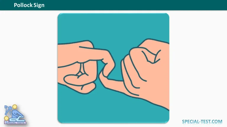
The Pollock Sign is a clinical examination test used to assess the function of the flexor digitorum profundus (FDP) muscle of the little finger in patients with suspected ulnar nerve injury. It helps clinicians identify weakness of distal finger flexion and provides useful information about the possible location of ulnar nerve dysfunction.
The test involves hooking the little fingers of both hands together and attempting to pull them apart while maintaining flexion. In patients with ulnar nerve dysfunction affecting the FDP muscle, the involved little finger may lose its flexed position, producing a positive Pollock Sign.
This simple physical examination maneuver can be used in orthopedic, neurological, and hand surgery assessments, particularly when evaluating proximal ulnar nerve injuries and cubital tunnel syndrome.
What Is the Purpose of the Pollock Sign?
The primary purpose of the Pollock Sign test is to evaluate the motor function of the flexor digitorum profundus muscle supplied by the ulnar nerve.
The FDP is a deep forearm muscle responsible for flexion of the distal interphalangeal (DIP) joints of the fingers. Its nerve supply is divided into two portions:
- Medial portion: Innervated by the ulnar nerve and primarily responsible for flexing the distal joints of the ring and little fingers.
- Lateral portion: Innervated by the anterior interosseous branch of the median nerve and primarily responsible for flexing the distal joints of the index and middle fingers.
Because the ulnar nerve supplies the medial FDP muscle in the forearm, proximal ulnar nerve injuries may cause weakness in little finger DIP flexion.
The Pollock Sign helps detect this motor deficit and contributes to distinguishing proximal ulnar nerve lesions from more distal injuries at the wrist.
How to Perform the Pollock Sign Test
The Pollock Sign is performed using the following steps:
- Patient positioning: Ask the patient to sit comfortably with both hands positioned in front of the body.
- Finger positioning: Instruct the patient to bend the little fingers of both hands, particularly at their distal interphalangeal joints, to create a hook-shaped position.
- Interlocking fingers: Ask the patient to hook the two little fingers together.
- Applying resistance: Instruct the patient to pull the hands apart while keeping the little fingers hooked and the distal phalanges flexed.
- Observing the result: Examine whether both little fingers maintain their flexed positions or whether one finger begins to straighten under resistance.
The examiner should compare the performance of both hands, paying particular attention to the ability to maintain distal interphalangeal joint flexion.
The movement should be performed gently, especially in patients with pain, recent trauma, or suspected tendon injury.
What Is a Positive Pollock Sign?
A positive Pollock Sign occurs when the patient cannot maintain flexion of the affected little finger while attempting to pull the hooked fingers apart.
The characteristic findings include:
- Extension of the distal phalanx of the affected little finger.
- Possible accompanying extension of the middle phalanx.
- Loss of the hooked finger position during resistance.
- Reduced ability to maintain DIP joint flexion compared with the unaffected hand.
These findings suggest weakness or paralysis of the FDP muscle, potentially resulting from ulnar nerve dysfunction.
Negative Pollock Sign
A negative Pollock Sign occurs when both little fingers maintain their hooked positions and resist the pulling force without straightening.
This finding suggests preserved FDP function during the maneuver. However, a negative test does not completely exclude partial ulnar nerve injury or mild neuropathy.
Clinical Significance of the Pollock Sign
The Pollock Sign is particularly relevant when evaluating the anatomical level of an ulnar nerve lesion.
1. Proximal Ulnar Nerve Injury
Proximal ulnar nerve injuries, including lesions around the elbow or proximal forearm, may impair the nerve branches supplying the medial FDP muscle.
Consequently, patients may demonstrate weakness of distal flexion in the little and ring fingers.
A positive Pollock Sign may therefore indicate injury at or proximal to the ulnar nerve branches supplying the FDP muscle.
2. Distal Ulnar Nerve Injury
Distal ulnar nerve lesions, particularly those occurring within Guyon’s canal at the wrist, generally spare the FDP muscle because its motor branches arise more proximally in the forearm.
Patients with these lesions may demonstrate weakness of intrinsic hand muscles while retaining the ability to flex the distal joint of the little finger.
This anatomical difference makes FDP testing useful when distinguishing proximal from distal ulnar nerve lesions.
3. Cubital Tunnel Syndrome
Cubital tunnel syndrome results from compression of the ulnar nerve at the elbow.
Affected individuals may develop numbness and tingling in the little finger and ulnar half of the ring finger, reduced hand strength, and impaired fine motor coordination.
In more advanced cases, weakness of the ulnar-innervated FDP may become apparent during physical examination.
The Pollock Sign can contribute to the clinical evaluation of these patients, although it is not sufficient to establish the diagnosis independently.
Other Clinical Tests for Ulnar Nerve Injury
The Pollock Sign should be interpreted alongside other physical examination findings.
Froment Sign: Assesses weakness of the adductor pollicis muscle. A positive result produces compensatory flexion of the thumb interphalangeal joint when the patient attempts to hold a piece of paper between the thumb and index finger.
Wartenberg Sign: Assesses abnormal abduction of the little finger due to weakness of ulnar-innervated hand muscles.
Finger Abduction and Adduction Testing: Evaluates the dorsal and palmar interosseous muscles, which are supplied by the ulnar nerve.
Isolated FDP Testing: The examiner stabilizes the proximal interphalangeal joint and asks the patient to actively flex the distal joint of the little finger, allowing more direct assessment of FDP function.
Together, these examinations help determine the distribution and severity of motor dysfunction.
Limitations of the Pollock Sign
Although useful as a bedside examination maneuver, the Pollock Sign has several limitations.
The test requires adequate patient cooperation and an understanding of the instructions. Pain, joint stiffness, limited mobility, and previous finger injuries may interfere with performance.
Importantly, weakness of distal finger flexion is not exclusive to ulnar nerve injury. Flexor tendon rupture or other musculoskeletal abnormalities may produce similar findings.
The sensitivity and specificity of the Pollock Sign have not been established adequately in the cited literature. Therefore, it should not be used as a standalone diagnostic test.
When clinically indicated, additional investigations such as nerve conduction studies, electromyography, and ultrasound may help confirm ulnar neuropathy and identify the site of injury.
Frequently Asked Questions (FAQs)
1. What does the Pollock Sign test?
The Pollock Sign evaluates the motor function of the flexor digitorum profundus muscle of the little finger, particularly its ability to maintain distal interphalangeal joint flexion against resistance.
2. Which nerve is assessed by the Pollock Sign?
The test primarily assesses the ulnar nerve motor supply to the medial portion of the flexor digitorum profundus muscle.
3. What indicates a positive Pollock Sign?
A positive result occurs when the affected little finger straightens during the pulling maneuver because the patient cannot maintain adequate flexion at the distal interphalangeal joint.
4. Does a positive Pollock Sign confirm ulnar nerve injury?
No. A positive result suggests FDP weakness, which may occur with proximal ulnar nerve dysfunction. However, tendon injuries, pain, and mechanical restrictions must also be considered.
5. Can the Pollock Sign help distinguish proximal and distal ulnar nerve injuries?
Yes. Weakness of the ulnar-innervated FDP suggests a lesion involving its motor supply in the forearm or more proximally. Isolated ulnar nerve injuries at the wrist generally preserve FDP function.
6. What is the difference between the Pollock Sign and Froment Sign?
The Pollock Sign evaluates FDP function in the little finger, whereas the Froment Sign evaluates weakness of the adductor pollicis muscle during a resisted pinch maneuver.
7. Is the Pollock Sign useful in cubital tunnel syndrome?
It may be useful as part of the motor examination, particularly when assessing suspected proximal ulnar nerve dysfunction. It should be combined with sensory examination, other motor tests, and electrodiagnostic investigations when appropriate.
Key Takeaway
The Pollock Sign is a simple clinical test for assessing flexor digitorum profundus weakness associated with ulnar nerve injury. It is performed by hooking both little fingers together and asking the patient to pull them apart while maintaining distal finger flexion.
Extension of the affected little finger during resistance represents a positive finding and may indicate impaired ulnar nerve motor function.
Its principal clinical value lies in assessing FDP weakness and supporting the localization of suspected ulnar nerve lesions. The result must always be interpreted in the context of the complete clinical examination.
References
- OrthoFixar. Pollock Sign: Clinical Examination, Procedure, and Interpretation. Read on OrthoFixar
- Goloborod’ko SA. New clinical motor test for cubital tunnel syndrome. Journal of Hand Therapy. 2012;25(4):422–423. PubMed – PMID: 22652328
- Dy CJ, Mackinnon SE. Ulnar neuropathy: evaluation and management. Current Reviews in Musculoskeletal Medicine. 2016;9(2):178–184. NCBI PubMed Central
- StatPearls. Anatomy, Shoulder and Upper Limb, Hand Compartments. NCBI Bookshelf. Read on NCBI
- StatPearls. Electrodiagnostic Evaluation of Ulnar Neuropathy. NCBI Bookshelf. Read on NCBI
- StatPearls. Flexor Tendon Lacerations. NCBI Bookshelf. Read on NCBI