Vertebral Artery Test (De Klyn Test): Purpose, Procedure, Positive Signs & Clinical Accuracy
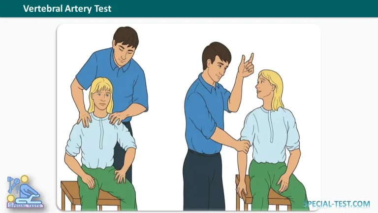
The Vertebral Artery Test, also called the De Klyn Test, is a traditional orthopedic examination used to investigate symptoms potentially associated with vertebrobasilar insufficiency (VBI). It involves positioning the cervical spine in rotation and extension while observing for neurological or vascular symptoms.
Historically, the test was used by physical therapists, orthopedic clinicians, and other healthcare professionals when assessing patients with neck pain, dizziness, or suspected vertebral artery compression, particularly before cervical spine manipulation.
However, contemporary research has questioned the diagnostic accuracy and safety of provocative vertebral artery testing. A negative Vertebral Artery Test cannot reliably exclude cervical vascular disease or establish that cervical manipulation is safe.
What Is the Vertebral Artery Test Used For?
The Vertebral Artery Test was traditionally designed to assess possible compromise of blood circulation through the vertebral arteries, which supply the posterior portion of the brain.
The vertebral arteries travel through the cervical spine and join to form the basilar artery. Together, these vessels supply structures including the brainstem, cerebellum, and parts of the occipital lobes.
Conditions affecting this circulation may cause neurological symptoms, particularly when blood flow becomes insufficient.
Historically, clinicians used the De Klyn Test to investigate:
- Vertebrobasilar insufficiency: Reduced blood supply to the posterior cerebral circulation.
- Vertebral artery compression: Potential mechanical narrowing of a vertebral artery during certain neck movements.
- Rotational vertebral artery syndrome: Also known as Bow Hunter syndrome, in which head rotation can produce dynamic vertebral artery occlusion.
- Cervical vascular symptoms: Dizziness, visual disturbances, or other neurological symptoms associated with neck movements.
Although older descriptions also associate the test with carotid artery abnormalities, it cannot reliably diagnose carotid, vertebral, or basilar artery stenosis.
Importantly, contemporary evidence does not support using the Vertebral Artery Test as a stand-alone vascular screening examination.
How to Perform the Vertebral Artery Test
The following describes the traditional clinical procedure for educational purposes, rather than a recommended routine screening technique. Because sustained cervical rotation and extension may provoke symptoms in patients with vascular disease, the maneuver should not be attempted independently.
1. Preliminary Patient Assessment
Before considering provocative cervical testing, a clinician should obtain a detailed medical history and assess the possibility of cervical vascular pathology.
Traditional preliminary assessment includes:
- Measurement of blood pressure.
- Evaluation of peripheral pulses in both arms.
- Examination of the carotid and subclavian arteries when clinically indicated.
- Assessment for vascular bruits.
- Screening for neurological abnormalities.
- Review of cardiovascular risk factors and previous vascular disorders.
Particular attention should be given to patients with recent severe neck pain or headache, visual disturbances, fainting, unexplained dizziness, or symptoms suggesting cervical artery dissection.
Abnormal findings require appropriate medical evaluation rather than provocative testing. Normal blood pressure and pulses do not exclude vertebral artery disease.
2. Seated Vertebral Artery Test
In the traditional seated technique:
- The patient sits upright on an examination chair.
- The examiner assesses the patient’s baseline symptoms.
- The cervical spine is rotated toward one side while the neck is extended.
- The position is historically maintained for approximately 20–30 seconds.
- The examiner observes for dizziness, nystagmus, visual symptoms, nausea, or other neurological changes.
- Older protocols describe repeating the maneuver toward the opposite side.
The purpose was to reproduce symptoms potentially associated with compromised posterior circulation.
Clinical caution: This provocative procedure is not recommended as a reliable method for clearing patients before cervical manipulation.
3. Supine Vertebral Artery Test (De Klyn Position)
Another traditional variation uses the supine position.
In this method, the patient lies on an examination table while the examiner supports the head near the edge of the table.
The historical De Klyn position combines cervical extension with head rotation. Older examination protocols describe maintaining this position for approximately 20–30 seconds while monitoring symptoms. Some descriptions additionally ask the patient to count aloud so that the examiner can observe speech changes.
This technique was intended to identify symptoms potentially associated with vascular compromise during cervical positioning.
However, symptom provocation does not directly measure arterial blood flow, and the maneuver cannot confirm or exclude vascular stenosis.
What Is a Positive Vertebral Artery Test?
Traditionally, a positive Vertebral Artery Test refers to the appearance or worsening of concerning symptoms during cervical extension and rotation.
Possible findings include:
- Vertigo: A sensation of spinning or abnormal movement.
- Nystagmus: Involuntary rhythmic eye movements.
- Visual disturbances: Blurred vision, double vision, or temporary visual changes.
- Nausea: Particularly when associated with dizziness or neurological symptoms.
- Dysarthria: Difficulty producing clear speech.
- Ataxia: Impaired balance or coordination.
- Presyncope: Feeling faint or close to losing consciousness.
- Other neurological symptoms: New weakness, sensory changes, or difficulty swallowing.
These symptoms may indicate a potentially serious condition and should prompt an appropriate medical assessment.
Nevertheless, a positive test does not establish a diagnosis of vertebral artery stenosis or vertebrobasilar insufficiency. Similar findings can occur with vestibular disorders and other neurological conditions.
If concerning symptoms arise during cervical examination, the maneuver should be stopped and the patient assessed for urgent medical referral.
How to Interpret Vertebral Artery Test Results
Positive Test Result
A positive response indicates that symptoms were provoked during cervical positioning.
The clinician should consider vascular, neurological, vestibular, and other possible causes rather than assuming vertebral artery compression has occurred.
When posterior circulation ischemia is suspected, urgent medical assessment may be necessary.
Negative Test Result
A negative result means that the maneuver did not reproduce the symptoms being observed.
However, the absence of symptoms does not establish normal vertebral artery blood flow.
A systematic review of premanipulative vertebrobasilar insufficiency tests reported sensitivity ranging from 0% to 57% across four studies of questionable methodological quality.
This low and inconsistent sensitivity means that patients with vascular abnormalities may produce false-negative findings.
Consequently, a negative result must never be interpreted as proof that cervical spine manipulation is safe.
Diagnostic Accuracy and Reliability
The clinical usefulness of the Vertebral Artery Test has been debated extensively.
A systematic review published in Manual Therapy examined the diagnostic accuracy of premanipulative vertebrobasilar insufficiency tests. The review found:
- Sensitivity ranging from 0% to 57%.
- Specificity ranging from 67% to 100%.
- Considerable variation between studies.
- Insufficient evidence to support reliable premanipulative screening.
These values relate to a group of premanipulative tests, not exclusively to the De Klyn maneuver.
Other research has similarly found that extension-rotation testing cannot reliably identify patients with reduced vertebral artery blood flow.
More recent clinical literature argues against routine pre-treatment positional vascular testing because of its poor sensitivity and inability to predict serious complications.
The International IFOMPT Cervical Framework emphasizes clinical reasoning, patient history, identification of vascular warning signs, and individualized risk assessment before musculoskeletal intervention.
Precautions and Contraindications
Provocative cervical positioning should be avoided when cervical vascular pathology is suspected.
Warning signs and clinical circumstances that require particular attention include:
- Sudden, severe, or unusual headache or neck pain.
- Suspected cervical artery dissection.
- Recent neck trauma accompanied by concerning symptoms.
- New visual disturbances, speech abnormalities, or swallowing difficulties.
- Unexplained fainting or significant balance problems.
- Known vertebral artery disease or previous posterior circulation ischemic events.
The presence of vascular risk factors such as hypertension, smoking, diabetes, and atherosclerosis should also inform the overall clinical assessment.
Sudden neurological symptoms, especially when accompanied by severe headache, neck pain, difficulty walking, double vision, weakness, or speech disturbance, require emergency medical evaluation.
Vertebral Artery Test vs. Other Diagnostic Tests
The Vertebral Artery Test is not equivalent to diagnostic vascular imaging.
When clinicians suspect vertebrobasilar insufficiency or rotational vertebral artery compression, appropriate investigations may include:
| Diagnostic Method | Clinical Purpose |
|---|---|
| CT Angiography (CTA) | Evaluates arterial anatomy, narrowing, and possible vascular abnormalities. |
| Magnetic Resonance Angiography (MRA) | Provides noninvasive assessment of cervical and intracranial blood vessels. |
| Duplex Doppler Ultrasound | Assesses blood-flow characteristics in accessible arterial segments. |
| Digital Subtraction Angiography (DSA) | Provides detailed vascular imaging and may be used in selected complex cases. |
| Dynamic Rotational Angiography | May identify position-dependent vertebral artery compression in suspected Bow Hunter syndrome. |
The choice of imaging depends on the patient’s clinical presentation and the suspected vascular disorder.
Clinical Significance of the De Klyn Test
The Vertebral Artery Test remains relevant to orthopedic and physical therapy education because it illustrates the historical approach to identifying possible vascular compromise during neck movements.
However, its role in contemporary clinical practice is limited.
Healthcare professionals should prioritize:
- Detailed history-taking and identification of vascular warning signs.
- Appropriate neurological and cardiovascular assessment.
- Differential diagnosis of dizziness and neck pain.
- Timely referral when vascular disease is suspected.
- Evidence-based risk assessment before cervical spine treatment.
No single positional test can reliably determine whether a patient is free from cervical vascular pathology.
Frequently Asked Questions (FAQs)
1. What is the purpose of the Vertebral Artery Test?
The Vertebral Artery Test was traditionally used to investigate symptoms potentially associated with vertebrobasilar insufficiency or vertebral artery compression during neck rotation and extension. It is not considered a reliable stand-alone diagnostic or premanipulative screening test.
2. What is another name for the Vertebral Artery Test?
The Vertebral Artery Test is commonly called the De Klyn Test. It is also associated with traditional cervical extension-rotation or vertebrobasilar provocation testing.
3. What indicates a positive Vertebral Artery Test?
A traditionally positive result involves the appearance of dizziness, vertigo, nystagmus, visual disturbances, nausea, or other neurological symptoms during cervical positioning. These findings require clinical evaluation but do not independently confirm vascular insufficiency.
4. How long is the Vertebral Artery Test held?
Traditional descriptions recommend maintaining cervical rotation and extension for approximately 20–30 seconds. This duration is historical and should not be interpreted as a validated or recommended modern screening protocol.
5. Is the Vertebral Artery Test safe?
The test involves provocative cervical positioning and may be inappropriate in patients with suspected vascular pathology. Because it lacks reliable diagnostic accuracy, routine use as a vascular clearance test before cervical manipulation is not supported by contemporary evidence.
6. Can a negative Vertebral Artery Test rule out vertebrobasilar insufficiency?
No. A negative finding cannot reliably exclude vertebral artery stenosis, cervical artery dissection, or other vascular abnormalities. The available research documents poor and variable sensitivity.
7. What is the difference between the Vertebral Artery Test and the Dix-Hallpike Test?
The Vertebral Artery Test historically aimed to provoke symptoms potentially related to vascular compromise. The Dix-Hallpike Test, by contrast, is a positional examination used primarily to diagnose posterior canal benign paroxysmal positional vertigo (BPPV).
Although both involve head positioning, their purposes and diagnostic evidence differ.
8. What is the best test for vertebral artery compression?
There is no single best examination for every patient. CTA, MRA, and vascular ultrasound may help investigate suspected vascular disease. In rotational vertebral artery syndrome, dynamic rotational angiography is particularly important for identifying position-dependent compression.
Conclusion
The Vertebral Artery Test (De Klyn Test) is a traditional cervical spine examination historically used to investigate possible vertebrobasilar insufficiency during neck rotation and extension.
Although symptoms such as vertigo, nystagmus, nausea, and visual disturbances may arise during the maneuver, these findings are not specific to vertebral artery pathology.
Current evidence demonstrates that premanipulative vertebral artery tests have limited diagnostic accuracy and cannot reliably identify patients at risk of vascular complications.
For modern clinical practice, comprehensive history-taking, neurological examination, vascular risk assessment, and appropriate diagnostic imaging provide a more evidence-based approach to evaluating suspected cervical vascular disorders.
References
- OrthoFixar. Vertebral Artery Test: Procedure and Interpretation. Updated November 2025.
https://orthofixar.com/special-test/vertebral-artery-test/ - Hutting N, et al. Diagnostic Accuracy of Premanipulative Vertebrobasilar Insufficiency Tests: A Systematic Review. Manual Therapy. 2013.
https://pubmed.ncbi.nlm.nih.gov/23127991/ - Côté P, et al. The Validity of the Extension-Rotation Test as a Clinical Screening Procedure Before Neck Manipulation: A Secondary Analysis. 1996.
https://pubmed.ncbi.nlm.nih.gov/8728458/ - Hutting N, et al. Yes, We Should Abandon Pre-Treatment Positional Testing of the Cervical Spine. 2020.
https://pubmed.ncbi.nlm.nih.gov/32861354/ - Rushton A, et al. International Framework for Examination of the Cervical Region for Potential of Vascular Pathologies of the Neck Prior to Musculoskeletal Intervention: International IFOMPT Cervical Framework. Journal of Orthopaedic & Sports Physical Therapy. 2023.
https://pubmed.ncbi.nlm.nih.gov/36099171/ - Benjamin R, Lui F. Vertebrobasilar Insufficiency. StatPearls. NCBI Bookshelf. Updated December 2025.
https://www.ncbi.nlm.nih.gov/books/NBK482259/ - Davis DD, Munakomi S, Kane SM. Rotational Vertebral Artery Syndrome. StatPearls. NCBI Bookshelf.
https://www.ncbi.nlm.nih.gov/books/NBK559022/