Spine Range of Motion: Normal Values for Cervical, Thoracic & Lumbar Spine
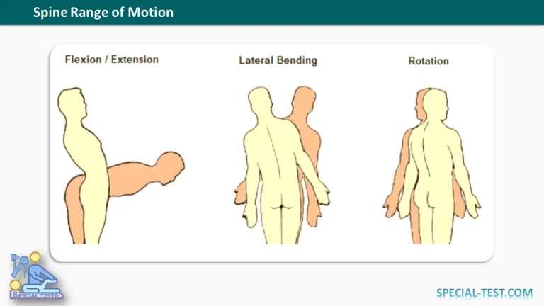
Spine range of motion (ROM) describes the degree of movement permitted by the vertebral column during flexion, extension, lateral bending, and axial rotation. Understanding normal spinal mobility is essential in orthopedic surgery, physical therapy, rehabilitation, and the clinical assessment of spinal disorders.
The human spine is divided into three major mobile regions: the cervical spine (C1–C7), thoracic spine (T1–T12), and lumbar spine (L1–L5). Each region has unique anatomical characteristics that determine its flexibility and movement patterns.
The cervical spine provides the greatest overall mobility, particularly during rotation and flexion-extension. The thoracic spine is relatively restricted by its articulation with the ribs, while the lumbar spine permits substantial flexion-extension but limited axial rotation. [1,2]
Normal Spine Range of Motion Chart
The following table summarizes approximate spinal range of motion values reported in biomechanical and radiographic research.
| Spinal Level | Flexion–Extension | Lateral Bending | Axial Rotation |
|---|---|---|---|
| Occiput–C1 (atlanto-occipital joint) | Approximately 18–26° | Approximately 3–6° to each side | Minimal, generally 0–3° to each side |
| C1–C2 (atlantoaxial joint) | Approximately 15–22° | Approximately 2–7° to each side | Approximately 30–40° to each side |
| C2–C7 (subaxial cervical spine) | Varies by individual segment | Major contributor to cervical lateral bending | Smaller contribution per level than C1–C2 |
| Thoracic spine (T1–T12) | Approximately 2–4° per segment | Approximately 2–4° per segment | Approximately 2–5° per segment |
| Lumbar spine (L1–S1) | Approximately 10–18° per segment | Varies by level; approximately 3–10° across the side-to-side arc | Approximately 2° per segment |
Important: These are approximate research-based values, not universal clinical reference limits. Different studies report different values depending on the testing method, patient characteristics, applied loading, and whether movement is measured in one direction or across the complete arc. Segmental measurements should not be confused with the total range of motion of an entire spinal region. [1–5]
Types of Spinal Movement
The vertebral column permits movement in three principal anatomical planes.
1. Flexion and Extension
Spinal flexion involves forward bending of the neck or trunk, whereas extension describes backward bending.
Flexion-extension occurs in the sagittal plane and is particularly important for activities such as looking downward, bending forward, standing upright, and reaching overhead.
The cervical and lumbar regions contribute substantially to sagittal movement, while thoracic mobility is comparatively restricted.
2. Lateral Bending
Lateral bending, also called lateral flexion, involves bending the neck or trunk toward either shoulder or side of the body.
This movement occurs primarily in the coronal plane and depends on the orientation of the facet joints, intervertebral discs, and surrounding ligamentous structures.
3. Axial Rotation
Axial rotation is the twisting movement of the spine around its vertical axis.
Rotation is especially prominent at the atlantoaxial joint (C1–C2). The thoracic spine also contributes to rotational movement, whereas the lumbar spine permits relatively little axial rotation because of its facet joint orientation and anatomical constraints.
Cervical Spine Range of Motion
The cervical spine is the most mobile part of the vertebral column. Its anatomy supports a wide range of head and neck movements.
Occiput–C1 (Atlanto-Occipital Joint)
The atlanto-occipital articulation connects the occipital bone of the skull to the atlas (C1).
Its primary movement is flexion and extension, producing the nodding motion commonly associated with saying “yes.”
Lateral bending is relatively limited, and axial rotation is minimal compared with the atlantoaxial joint.
A large biomechanical study published in 2025 reported a mean combined flexion-extension range of approximately 26.4° at the occiput–C1 segment, demonstrating that this articulation contributes significantly to upper cervical mobility. [1]
C1–C2 (Atlantoaxial Joint)
The atlantoaxial joint lies between the atlas (C1) and axis (C2).
This articulation is responsible for a substantial proportion of cervical rotation, enabling the head-turning movement associated with saying “no.”
The odontoid process, or dens, of C2 acts as a pivot around which C1 rotates.
In an in vivo MRI study, maximum C1–C2 axial rotation averaged approximately 36.2° to each side. Other biomechanical investigations have reported similar values, although measurements vary with the study method. [3]
C2–C7 (Subaxial Cervical Spine)
The subaxial cervical region extends from C2 to C7 and contributes substantially to neck flexion, extension, lateral bending, and rotation.
Movement is distributed unequally across individual motion segments. The middle and lower cervical levels, especially C4–C5 and C5–C6, frequently contribute significantly to flexion-extension.
A systematic review of cervical spine kinematics confirmed that cervical mobility is complex and cannot be accurately represented by assuming that every vertebral level moves through the same angle. [6]
Thoracic Spine Range of Motion
The thoracic spine consists of 12 vertebrae and forms the posterior attachment of the rib cage.
Compared with the cervical and lumbar spine, thoracic flexion-extension is generally more restricted because of the rib cage, facet joint orientation, and surrounding ligamentous structures.
However, the thoracic spine plays an important role in trunk rotation and lateral bending.
A systematic review examining segmental thoracic motion reported pooled mean ranges of approximately:
- Flexion-extension: 1.9–3.8° per segment.
- Lateral bending: 2.1–4.4° per segment.
- Axial rotation: 2.4–5.2° per segment.
These values depend on spinal level and experimental conditions. [4]
Thoracic range of motion is clinically relevant when evaluating spinal deformities, postural abnormalities, thoracic stiffness, and functional limitations.
Lumbar Spine Range of Motion
The lumbar spine consists of five large vertebrae designed primarily for weight-bearing and stability.
The lumbar region permits substantial flexion and extension, which are essential for bending forward, lifting, and returning to an upright position.
Its capacity for axial rotation is considerably lower than that of the cervical and thoracic regions.
Radiographic research in asymptomatic individuals has demonstrated approximately 14° of combined flexion-extension and 2° of axial rotation at individual lumbar motion segments. Segmental lateral bending varies, with some upper lumbar levels demonstrating greater mobility than the lumbosacral junction. [5]
These anatomical characteristics help explain why excessive twisting combined with loading may place considerable mechanical demands on the lumbar spine.
Factors Affecting Normal Spinal Range of Motion
Spinal mobility varies between individuals and is influenced by several factors:
- Age: Cervical and lumbar flexibility generally decline with advancing age.
- Degenerative changes: Disc degeneration, facet joint osteoarthritis, and other structural changes may reduce mobility.
- Muscle flexibility: Restricted muscles and soft tissues can limit active spinal movement.
- Pain and muscle guarding: Pain may temporarily restrict spinal motion without necessarily indicating a fixed structural limitation.
- Previous spinal surgery: Spinal fusion reduces motion at the fused levels and may alter the distribution of movement across adjacent segments.
- Measurement technique: Active versus passive movement, patient positioning, and the instruments used can influence recorded ROM values.
A systematic review published in PubMed identified age-related reductions in lumbar flexion, extension, and lateral bending, emphasizing the importance of age-appropriate interpretation. [7]
Clinical Assessment of Spine Range of Motion
Measuring spinal ROM is an important part of the musculoskeletal examination.
Clinicians may use a goniometer, inclinometer, cervical range-of-motion device, or other validated measurement systems to quantify movement.
The examination generally includes observing active flexion, extension, right and left lateral bending, and right and left rotation.
When clinically indicated, imaging and specialized functional assessments may provide additional information about individual vertebral motion segments.
A systematic review found evidence supporting the reliability and validity of several cervical ROM measurement devices, including the cervical range-of-motion instrument and single inclinometer. [8]
Importantly, reduced spinal range of motion alone does not establish a diagnosis. Findings should be interpreted alongside symptoms, neurological examination, functional limitations, and other clinical information.
Frequently Asked Questions (FAQ)
1. What is the normal range of motion of the spine?
Normal spinal ROM depends on the region being examined, the individual’s age, and the measurement method. The cervical spine is generally the most mobile region, the thoracic spine has restricted flexion-extension, and the lumbar spine permits substantial forward and backward bending with limited rotation.
2. Which spinal segment has the greatest rotational movement?
The C1–C2 atlantoaxial joint has the greatest individual segmental axial rotation. It contributes a large proportion of overall cervical rotation, with approximately 30–40° of rotation to each side reported in several studies.
3. Which part of the spine allows the most flexion and extension?
The cervical and lumbar regions permit substantial flexion-extension. Within the upper cervical spine, the occiput–C1 articulation is an important contributor to flexion-extension, while the lower cervical segments contribute significantly to total neck movement.
4. Why is lumbar spine rotation limited?
Lumbar axial rotation is restricted primarily by the orientation of the facet joints and associated spinal structures. This anatomical arrangement favors flexion-extension and stability while limiting twisting at individual lumbar levels.
5. Does spinal range of motion decrease with age?
Yes. Research demonstrates that cervical and lumbar ROM generally decreases with age, although the extent of reduction differs between movement planes and individuals. Normal aging should be considered when interpreting spinal mobility measurements.
6. How is spinal range of motion measured?
Spinal ROM can be assessed through clinical examination using goniometers, inclinometers, specialized cervical measurement devices, and other validated tools. Radiographic or advanced imaging techniques may be used when a more detailed evaluation of segmental motion is clinically necessary.
7. Is reduced spine range of motion always abnormal?
No. Reduced mobility may reflect normal age-related changes, pain, muscle tightness, or structural limitations. A clinically meaningful assessment considers the patient’s symptoms, baseline mobility, neurological findings, and functional status rather than relying solely on a numerical ROM value.
Conclusion
Understanding spine range of motion is fundamental to orthopedic examination, spinal biomechanics, and rehabilitation.
The cervical spine provides substantial multidirectional mobility, with the atlantoaxial joint contributing prominently to rotation. The thoracic spine balances mobility with rib cage stability, while the lumbar spine is adapted for weight-bearing and flexion-extension with restricted axial rotation.
Although approximate ROM charts provide useful educational guidance, normal spinal movement varies across individuals and individual vertebral levels. Evidence-based interpretation requires consideration of anatomical differences, age, measurement techniques, and the patient’s clinical presentation.
References
- Farber SH, et al. Clinical effect of occipitocervical and subaxial cervical fusion constructs on range of motion: comprehensive guide based on biomechanical cadaveric testing on 1009 motion segments. Journal of Neurosurgery: Spine. 2025. PubMed
- Range of Motion and Neutral Zone of All Human Spinal Motion Segments: A Data Collection of 30 Years of In Vitro Experiments Performed Under Standardized Testing Conditions. 2025. PubMed
- Kinematics of the upper cervical spine in rotation: in vivo three-dimensional analysis. Spine. 2004. PubMed
- Challenging the Conventional Standard for Thoracic Spine Range of Motion: A Systematic Review. 2016. PubMed
- Pearcy MJ. Stereo radiography of lumbar spine motion. Acta Orthopaedica Scandinavica Supplementum. 1985. PubMed
- Kinematics of the Cervical Spine Under Healthy and Degenerative Conditions: A Systematic Review. 2022. PubMed
- Intolo P, et al. The effect of age on lumbar range of motion: a systematic review. Manual Therapy. 2009. PubMed
- Williams MA, et al. A systematic review of reliability and validity studies of methods for measuring active and passive cervical range of motion. Journal of Manipulative and Physiological Therapeutics. 2010. PubMed