Schober Sign: Definition, Purpose, Procedure, and Positive Results
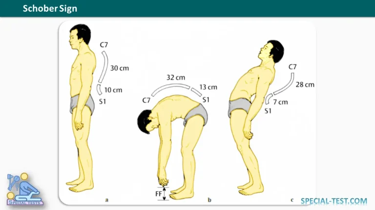
The Schober Sign (Schober Test) is a clinical examination technique used to assess lumbar spine mobility, particularly during forward flexion. It is commonly used when evaluating patients with suspected ankylosing spondylitis and other conditions that may cause restricted movement of the lumbar spine.
This simple, noninvasive test measures the change in distance between specific skin markings over the lower back as the patient bends forward. Reduced movement between these markings may indicate limited lumbar flexion and can help clinicians identify spinal stiffness associated with inflammatory rheumatic diseases.
Although the Schober Sign is useful during musculoskeletal assessment, it should not be used alone to diagnose ankylosing spondylitis. Its findings must be interpreted alongside the patient’s symptoms, physical examination, laboratory investigations, and imaging when indicated.
What Is the Schober Sign Used For?
The Schober Sign is primarily used to measure lumbar spine mobility, especially the range of motion during forward flexion.
Its main clinical applications include:
- Assessment of lumbar flexion: Evaluates how much the skin markings over the lumbar spine separate when the patient bends forward.
- Screening for restricted spinal mobility: Helps identify reduced lumbar movement in patients with back stiffness.
- Evaluation of suspected ankylosing spondylitis: Supports the clinical examination of patients with suspected axial spondyloarthritis.
- Monitoring spinal mobility: May be used to document changes in lumbar flexion during follow-up examinations.
- Assessment of inflammatory spinal disease: Can help detect functional limitation associated with inflammatory conditions affecting the spine.
The test is particularly relevant in ankylosing spondylitis, a chronic inflammatory disease that primarily affects the axial skeleton and may lead to progressive spinal stiffness and reduced mobility.
However, restricted lumbar flexion is not specific to ankylosing spondylitis. Other musculoskeletal conditions, including degenerative spinal disease, pain-related muscle guarding, and mechanical restrictions, may also reduce the measured range of motion.
How to Perform the Schober Sign
The Schober Test is performed with the patient standing and requires a measuring tape and a skin-safe marker.
Patient Position
Ask the patient to stand upright with the knees extended and the lumbar spine in a neutral position. Ensure that the patient is comfortable and can bend forward safely.
Identify the Anatomical Landmarks
The examiner identifies the lumbosacral junction, traditionally using the level of the S1 spinous process or an equivalent landmark described by the specific test protocol.
A skin mark is placed at the selected landmark. A second mark is then made 10 cm superior to the first mark.
Accurate identification of the landmarks is important because differences in marking technique can affect the measurement.
Measure Lumbar Flexion
Ask the patient to bend forward as far as possible while keeping the knees extended, provided this can be done comfortably.
As the lumbar spine flexes, the distance between the two skin markings increases. Measure and record the distance between the marks at maximum forward flexion.
Compare the initial and final measurements to determine the change in distance.
Interpret the Measurement
In the traditional Schober Test, the initial 10 cm distance is expected to increase during forward flexion. The amount of separation provides an estimate of lumbar mobility.
The modified Schober Test uses different marking landmarks and distances. For this reason, the examiner should follow one standardized method and use its corresponding reference values rather than mixing measurements from different versions.
What Is a Positive Schober Sign?
A positive Schober Sign generally indicates reduced lumbar spine flexion when the distance between the skin markings increases less than expected during forward bending.
The traditional Schober Test commonly uses an increase from 10 cm to approximately 15 cm as a reference for normal mobility, although expected values vary with the method used and the individual being examined.
A smaller increase suggests restricted lumbar flexion. For example, if the distance increases from 10 cm to only 12 cm, the measured increase is 2 cm, which may indicate limited lumbar mobility.
The result should be interpreted using the relevant test protocol and clinical context rather than a single universal cutoff.
What Causes a Positive Result?
Reduced lumbar flexion may occur in several conditions, including:
- Ankylosing spondylitis and other forms of axial spondyloarthritis.
- Degenerative changes affecting the lumbar spine.
- Painful lumbar conditions that limit forward bending.
- Spinal stiffness associated with structural or inflammatory disease.
- Reduced flexibility or other mechanical limitations of the lower back.
In ankylosing spondylitis, inflammation and subsequent structural changes can progressively restrict spinal movement. A reduced Schober measurement may therefore support the clinical suspicion of axial spondyloarthritis, particularly when accompanied by chronic inflammatory back pain and other characteristic findings.
Nevertheless, a positive result does not confirm the diagnosis, and a normal result does not completely exclude early inflammatory spinal disease.
Schober Test vs. Modified Schober Test
Several variations of the test are used in clinical practice. Understanding which method is being performed is essential for accurate documentation.
Traditional Schober Test
The traditional method begins with a 10 cm distance between the selected skin markings. The examiner measures how much this distance increases during maximum forward flexion.
The classic reference is an increase toward approximately 15 cm, representing an increase of about 5 cm.
Modified Schober Test
The modified Schober Test uses different anatomical landmarks, commonly including a mark at the lumbosacral junction and additional marks above and below it. The initial measurement and expected change differ from those of the traditional method.
The modified technique is widely used in clinical assessment because it aims to provide a more standardized estimate of lumbar flexion.
Clinical tip: Always document the version of the Schober Test used, the baseline distance, the final distance, and the change in centimeters. This makes the findings easier to interpret and compare during follow-up.
Clinical Significance and Limitations
The Schober Sign is useful because it is quick, inexpensive, and easy to perform without specialized equipment. It provides an objective measurement that can supplement the examination of patients with suspected inflammatory spinal disease.
However, several limitations should be considered:
- Skin movement does not perfectly represent movement between the vertebrae.
- Results may vary with landmark identification and examiner technique.
- Pain, muscle tightness, age, and body habitus can influence the measurement.
- The test assesses lumbar flexion rather than the entire range of spinal movement.
- It cannot independently distinguish inflammatory disease from degenerative or other mechanical causes of restricted mobility.
For suspected ankylosing spondylitis, clinicians should integrate the Schober Test with the patient’s history, spinal examination, chest expansion when appropriate, sacroiliac joint assessment, and further investigations when clinically indicated.
Key Points
- The Schober Sign assesses lumbar spine mobility during forward flexion.
- It is commonly used when evaluating suspected ankylosing spondylitis and other causes of spinal stiffness.
- In the traditional test, the distance between two markings starts at 10 cm and should increase during forward bending.
- A smaller-than-expected increase suggests restricted lumbar flexion.
- The test supports clinical assessment but cannot independently diagnose or exclude ankylosing spondylitis.
Frequently Asked Questions (FAQ)
1. What does the Schober Sign test for?
The Schober Sign measures lumbar spine mobility during forward flexion. It is particularly useful for identifying restricted lumbar movement in patients with suspected ankylosing spondylitis or other spinal disorders.
2. How is the Schober Test performed?
The patient stands upright while the examiner marks a point near the lumbosacral junction and another point 10 cm above it. The patient then bends forward, and the examiner measures the increase in distance between the markings.
3. What is a positive Schober Sign?
A positive result generally indicates reduced lumbar flexion, demonstrated by a smaller-than-expected increase in the distance between the markings during forward bending.
4. What is the normal Schober Test measurement?
In the traditional Schober Test, a baseline distance of 10 cm is expected to increase to approximately 15 cm during forward flexion. Normal values and cutoffs may differ according to the test variant and clinical protocol.
5. Is the Schober Test used to diagnose ankylosing spondylitis?
No. The test helps assess spinal mobility and may support the evaluation of ankylosing spondylitis, but it is not diagnostic by itself. Diagnosis requires consideration of clinical features and, when appropriate, laboratory tests and imaging.
6. What is the difference between the Schober Test and the modified Schober Test?
The two techniques use different marking methods and measurement protocols. The modified Schober Test incorporates additional landmarks, and its measurements should be interpreted using the reference criteria established for that version.
7. Can the Schober Test be normal in ankylosing spondylitis?
Yes. Lumbar mobility may remain relatively preserved in early disease or in patients without substantial spinal restriction. A normal result does not exclude ankylosing spondylitis.
References
- Clinical Tests for the Musculoskeletal System, 3rd edition.
- Sieper J, Poddubnyy D. Axial spondyloarthritis. The Lancet. 2017;390(10089):73–84.
- Assessment of lumbar mobility and spinal function in axial spondyloarthritis. Relevant clinical literature indexed in PubMed and available through the National Center for Biotechnology Information (NCBI).