Derbolowsky Test: Procedure, Positive Findings, Interpretation & Clinical Significance
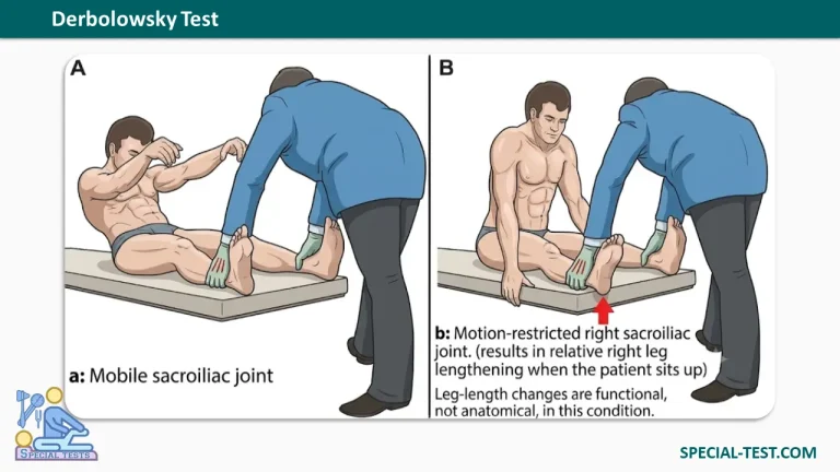
The Derbolowsky Test, also known as the supine-to-sit test or long sitting test, is a clinical orthopedic examination used to assess changes in apparent leg length during movement from a lying to a sitting position. It is traditionally performed to identify possible pelvic asymmetry and sacroiliac joint (SIJ) movement restrictions.
The test involves comparing the positions of the medial malleoli (the bony prominences on the inner sides of the ankles) while the patient is supine and after sitting upright. A change in the relative position of the ankles may suggest a functional leg length discrepancy or altered lumbopelvic mechanics.
Although commonly described in manual medicine, the Derbolowsky Test has limited scientific validation as an independent diagnostic method. Its findings should be interpreted alongside the patient’s symptoms, medical history, and other physical examination results.
What Is the Derbolowsky Test Used For?
The primary purpose of the Derbolowsky Test is to identify changes in apparent leg length associated with movement of the pelvis and lower extremities.
Traditionally, clinicians use the test to evaluate:
- Apparent or functional leg length discrepancies.
- Possible sacroiliac joint movement restrictions.
- Pelvic rotation or asymmetrical pelvic positioning.
- Changes in lower-limb alignment during trunk movement.
- Muscular tightness that may influence pelvic mechanics.
The test does not directly measure the anatomical length of the femur or tibia. Instead, it evaluates whether the relative positions of the ankles change during a transition from lying down to sitting.
How to Perform the Derbolowsky Test
The Derbolowsky Test is performed with the patient initially lying supine on an examination table. The examiner compares the positions of both medial malleoli before and after the patient sits upright.
Step-by-Step Procedure
- Patient positioning: Ask the patient to lie comfortably in the supine position with both lower extremities extended and positioned symmetrically.
- Examiner positioning: Stand at the foot of the examination table and gently grasp both ankles.
- Initial assessment: Place the thumbs over the medial malleoli of both ankles. Compare their relative levels and rotational positions to identify any apparent leg length difference.
- Sitting movement: Instruct the patient to move from the supine position into a long sitting position. The examiner may assist if necessary, or the patient may use their hands for support.
- Avoid interference: Ensure that the lower extremities can move freely without significant friction or obstruction from the examination table.
- Reassessment: Once the patient is sitting upright with the knees extended, compare the medial malleoli again. Observe whether one ankle appears to advance farther than the other.
- Forward trunk flexion: Ask the patient to bend the trunk forward toward the extended knees as far as comfortably possible, without forcing the movement.
- Repeat the examination: Perform the test several times to assess the consistency of the findings and reduce the potential influence of muscular tension or inconsistent positioning.
The examiner should avoid excessive traction on the ankles and document any discomfort, asymmetrical movement, or changes in apparent leg length.
What Is a Positive Derbolowsky Test?
A positive Derbolowsky Test is traditionally characterized by a change in the relative position of the medial malleoli when the patient moves from the supine to the sitting position.
In the traditional interpretation, the leg on the side of a restricted sacroiliac joint may appear relatively shorter or equal in length while the patient is supine but advance and appear longer when the patient sits upright.
This phenomenon is sometimes described as apparent leg lengthening or the advancement phenomenon.
For example, if both medial malleoli are initially aligned while the patient is lying down, but the right medial malleolus moves farther forward during sitting, the examiner may record a positive advancement phenomenon on the right side.
Historically, this finding has been interpreted as suggesting altered pelvic rotation or restricted sacroiliac joint movement.
However, a positive result does not confirm sacroiliac joint dysfunction, pelvic torsion, or anatomical leg length discrepancy. Similar changes may occur because of differences in positioning, muscle tension, or other musculoskeletal factors.
Clinical Interpretation and Diagnostic Significance
Traditional descriptions of the Derbolowsky Test consider an apparent difference of approximately 1–2 cm between the medial malleoli to be potentially clinically significant.
However, this threshold should be regarded as a historical interpretation rather than a scientifically validated diagnostic cutoff.
Several additional findings require consideration:
- A larger apparent discrepancy with muscular pain: May prompt evaluation for hamstring tightness, muscular shortening, or altered pelvic positioning.
- Apparent leg length changes with limited trunk flexion: May suggest muscle tightness or restrictions involving the hips, pelvis, or lumbar spine.
- Pain during the test: May be associated with musculoskeletal disorders or, when accompanied by neurological symptoms, lumbar nerve involvement.
- Marked asymmetry with radiating leg pain: Requires additional evaluation for lumbar spine disorders, including possible nerve root irritation.
Traditional descriptions also mention apparent differences exceeding 5 cm in association with radicular symptoms and compensatory knee flexion. However, this finding is not a validated diagnostic criterion for lumbar disc herniation and should not be interpreted in isolation.
Differential Diagnosis of a Positive Derbolowsky Test
A change in apparent leg length does not necessarily indicate sacroiliac joint pathology. Several conditions can influence lower-limb alignment and create similar examination findings.
1. Hamstring Tightness
Shortened or tight hamstring muscles can restrict pelvic movement during forward trunk flexion. This may influence the apparent position of the ankles and contribute to inconsistent test findings.
2. Anatomical Leg Length Discrepancy
A true or structural leg length discrepancy occurs when the bones of one lower extremity differ in length from those of the opposite side.
Unlike an apparent discrepancy related to positioning or muscular factors, structural differences cannot be reliably established through the Derbolowsky Test alone.
3. Hip Contractures
Hip abduction or adduction contractures may cause compensatory pelvic tilt and alter apparent leg length.
Contractures involving the hip or knee should therefore be considered when interpreting the examination.
4. Lumbar Spine Disorders
Lumbar disc pathology, nerve root irritation, or other spinal conditions may produce low back pain, restricted movement, or radiating lower-extremity symptoms.
These disorders require appropriate neurological and musculoskeletal examination rather than diagnosis based solely on ankle alignment.
5. Functional Pelvic Asymmetry
Changes in posture, muscular tension, or pelvic positioning can alter the apparent relationship between the lower extremities without representing a true difference in bone length.
Reliability and Limitations of the Derbolowsky Test
The diagnostic reliability of the Derbolowsky Test remains uncertain.
Research examining related sacroiliac joint mobility tests has identified limitations in examiner agreement and diagnostic validity. A study investigating the supine-to-sit test and other pelvic mobility assessments reported low sensitivity for identifying innominate torsion.
Furthermore, a systematic review of sacroiliac joint mobility tests found insufficient evidence to establish the validity of several commonly used palpation-based assessments.
Consequently, the Derbolowsky Test should not be used as the sole basis for diagnosing sacroiliac joint dysfunction or determining treatment.
When true leg length discrepancy is suspected, additional assessment methods may include:
- Standing block testing to assess pelvic leveling.
- Direct clinical leg length measurements.
- Assessment of hip and knee range of motion.
- Full-length standing radiographs when clinically indicated.
A systematic review published in PLOS ONE found that the standing block test appeared to be the most useful clinical assessment for leg length discrepancy, while full-length standing anteroposterior radiography provided a reliable method of assessing anatomical differences.
Clinical Importance of the Derbolowsky Test
The Derbolowsky Test remains a traditional component of some manual medicine and musculoskeletal examination approaches.
Its principal value is as an observational assessment of changes in apparent lower-limb alignment during movement.
However, clinicians should interpret its findings cautiously and integrate them with a comprehensive physical examination. Pain provocation tests, neurological examination, hip assessment, and appropriate leg length measurement techniques may provide additional information when investigating low back pain or pelvic dysfunction.
Frequently Asked Questions (FAQ)
What is the Derbolowsky Test?
The Derbolowsky Test is a clinical examination that compares apparent leg length in supine and sitting positions. It is traditionally used to investigate possible pelvic asymmetry and sacroiliac joint movement restrictions.
What does a positive Derbolowsky Test indicate?
A positive result indicates a change in the relative position of the medial malleoli when the patient moves from lying down to sitting. Although traditionally associated with pelvic rotation or sacroiliac joint restriction, it cannot independently confirm these conditions.
How is the Derbolowsky Test performed?
The patient lies supine with both legs extended. The examiner compares the levels of the medial malleoli and repeats the comparison after the patient sits upright. Any change in relative ankle alignment is recorded.
Is the Derbolowsky Test reliable?
The Derbolowsky Test has limited direct scientific validation. Studies of related sacroiliac joint mobility assessments have demonstrated inconsistent diagnostic performance. Therefore, its results should be interpreted alongside other clinical findings.
What is the difference between the Derbolowsky Test and the supine-to-sit test?
The terms are often used interchangeably. Both examinations involve comparing apparent leg length while the patient transitions from a supine to a sitting position.
Can the Derbolowsky Test diagnose sacroiliac joint dysfunction?
No. A positive test alone cannot establish sacroiliac joint dysfunction. Diagnosis requires consideration of the patient’s symptoms, physical examination findings, and other appropriate investigations.
What causes an apparent leg length discrepancy?
Possible causes include pelvic positioning, hamstring tightness, hip or knee contractures, postural adaptations, and other musculoskeletal factors. A structural leg length difference must be distinguished from these functional influences.
Can the Derbolowsky Test diagnose a herniated lumbar disc?
No. A herniated disc cannot be diagnosed using this test. Radiating leg pain, numbness, weakness, or other neurological findings require appropriate clinical assessment and, when indicated, additional investigations.
References
- Orthofixar – Derbolowsky Test: Procedure, Positive Findings, and Leg Length Assessment.
- Levangie PK. Four Clinical Tests of Sacroiliac Joint Dysfunction: Association With Innominate Torsion. Physical Therapy. 1999.
- Validity and Reliability of Palpatory Clinical Tests of Sacroiliac Joint Mobility: A Systematic Review and Meta-analysis. 2021.
- Alfuth M, et al. Leg Length Discrepancy: A Systematic Review on the Validity and Reliability of Clinical Assessments and Imaging Diagnostics. PLOS ONE. 2021.
- Saueressig T, et al. Diagnostic Accuracy of Clusters of Pain Provocation Tests for Detecting Sacroiliac Joint Pain: Systematic Review With Meta-analysis. 2021.