Beevor Sign: How to Perform, Positive Test & Clinical Significance
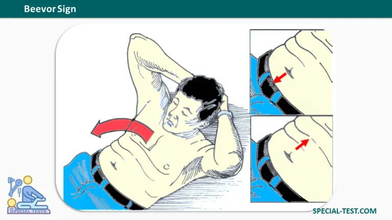
Beevor Sign (Beevor’s sign) is a clinical neurological examination finding used to identify weakness or paralysis of the rectus abdominis muscle, particularly its lower portion. It is characterized by abnormal upward displacement of the umbilicus (belly button) when a patient attempts to flex the neck or sit up from a supine position.
The sign is traditionally associated with spinal cord lesions involving the T10–T12 spinal segments. However, it is also an important clinical finding in several neuromuscular disorders, particularly facioscapulohumeral muscular dystrophy (FSHD).
Beevor Sign was first described by British neurologist Charles Edward Beevor in 1898. It remains a useful bedside examination technique because it can reveal subtle abdominal muscle weakness without requiring specialized equipment.
Under normal circumstances, the upper and lower portions of the rectus abdominis contract together during trunk flexion, keeping the umbilicus relatively stationary. When the lower abdominal muscles are weaker than the upper muscles, the stronger upper fibers pull the umbilicus upward, producing a positive Beevor Sign.
What Is Beevor Sign Used For?
The primary purpose of Beevor Sign is to evaluate functional weakness of the abdominal musculature and identify possible neurological or neuromuscular disorders.
Its main clinical applications include:
- Spinal cord lesion localization: Helps identify possible lesions involving the lower thoracic spinal cord, especially the T10–T12 segments.
- Abdominal muscle assessment: Detects disproportionate weakness of the upper or lower rectus abdominis.
- Facioscapulohumeral muscular dystrophy screening: Provides an additional diagnostic clue in patients suspected of having FSHD.
- Neuromuscular examination: May reveal characteristic muscle involvement in certain inherited or acquired myopathies.
- Neurological assessment: Complements motor, sensory, reflex, and other examination findings when evaluating suspected spinal cord disease.
Importantly, Beevor Sign is a supportive clinical finding rather than a definitive diagnostic test. Its interpretation must be combined with the patient’s history, neurological examination, and appropriate investigations.
How to Perform the Beevor Sign Test
The Beevor Sign test is a simple bedside physical examination that assesses the movement of the umbilicus during abdominal muscle activation.
Patient Position
- Ask the patient to lie supine (face upward) on an examination table.
- Position the knees in slight flexion, with both feet resting flat on the examination surface.
- Ensure that the abdominal region is adequately exposed to observe the umbilicus.
- Ask the patient to relax the abdominal muscles before beginning.
Examination Procedure
Step 1: Observe the resting position of the umbilicus and assess the symmetry of the abdominal wall.
Step 2: Ask the patient to lift the head and shoulders slightly from the examination table, as though beginning a sit-up.
Step 3: The patient may keep the arms crossed over the chest to avoid using the upper extremities for assistance.
Step 4: Observe the position and direction of movement of the umbilicus during the contraction.
Step 5: Determine whether the umbilicus remains stationary or moves upward, downward, or laterally.
Step 6: Repeat the maneuver if necessary to confirm the finding.
Alternative maneuvers described in clinical examination references include resisted head raising, coughing, or bilateral leg elevation to assess abdominal muscle activation.
Normal Beevor Sign (Negative Test)
A negative Beevor Sign occurs when the umbilicus remains approximately in its original position during trunk flexion.
This indicates relatively balanced contraction of the upper and lower rectus abdominis muscles.
What Is a Positive Beevor Sign?
A positive Beevor Sign is characterized by upward movement of the umbilicus when the patient attempts to raise the head or sit up.
The upward displacement occurs because the lower portion of the rectus abdominis is weaker or paralyzed, allowing the stronger upper fibers to pull the umbilicus toward the chest.
The NCBI StatPearls reference describes upward movement exceeding 1 cm as a positive finding.
Interpretation of Umbilical Movement
| Umbilical movement | Clinical interpretation |
|---|---|
| No significant movement | Negative Beevor Sign; relatively balanced abdominal contraction |
| Upward displacement | Weakness of the lower rectus abdominis; classical positive Beevor Sign |
| Downward displacement | Weakness of the upper rectus abdominis; inverted Beevor Sign |
| Lateral displacement | Asymmetric abdominal muscle weakness, with movement toward the stronger side |
The direction of umbilical movement can help identify the distribution of abdominal muscle weakness, but it does not establish an exact spinal cord lesion level by itself.
Beevor Sign and Spinal Cord Lesions
Beevor Sign was historically described as an indicator of lower thoracic spinal cord dysfunction.
Spinal cord lesions affecting the T10–T12 segments can impair the motor supply to the lower portions of the abdominal musculature while preserving relatively stronger upper abdominal muscles.
Consequently, during an attempted sit-up, the intact upper rectus abdominis pulls the umbilicus upward.
Potential neurological causes include:
- Thoracic spinal cord tumors.
- Traumatic spinal cord injuries.
- Spinal cord infarction.
- Other focal spinal cord lesions affecting the relevant motor pathways.
An acute appearance of Beevor Sign has also been reported in patients with spinal cord infarction.
Although the finding may help neurological localization, clinicians must evaluate accompanying symptoms such as limb weakness, sensory changes, altered reflexes, and bladder dysfunction.
Sudden weakness or new bladder or bowel dysfunction associated with suspected spinal cord disease requires urgent medical evaluation.
Beevor Sign in Facioscapulohumeral Muscular Dystrophy (FSHD)
One of the most clinically important associations of Beevor Sign is facioscapulohumeral muscular dystrophy (FSHD).
FSHD is an inherited neuromuscular disorder characterized by progressive muscle weakness, commonly involving the facial muscles, shoulder girdle, upper arms, and trunk.
In some patients, the lower portion of the rectus abdominis becomes disproportionately weak, resulting in upward umbilical displacement during trunk flexion.
Beevor Sign can be observed even before obvious functional abdominal weakness develops.
Diagnostic Accuracy in FSHD
Early research suggested that Beevor Sign was highly sensitive and specific for FSHD.
A 1990 study involving 30 patients with FSHD reported a positive sign in 27 patients (90%), while none of the 40 patients in the comparison group demonstrated the finding.
However, later studies reported lower sensitivity.
In a 2010 study published in the Journal of Neurology, researchers examined 28 genetically confirmed FSHD patients and 65 individuals with other neuromuscular diseases.
The findings included:
- Beevor Sign was positive in 15 of 28 FSHD patients, approximately 54%.
- Only 2 of 65 patients without FSHD demonstrated a positive result.
- The sign was more frequent in patients with typical FSHD features than in those with atypical presentations.
These findings suggest that Beevor Sign may be relatively specific for FSHD in selected clinical populations, but its sensitivity varies.
Therefore, a negative Beevor Sign does not exclude FSHD, and a positive result cannot independently confirm the diagnosis.
Other Conditions Associated With a Positive Beevor Sign
Although Beevor Sign is classically associated with spinal cord lesions and FSHD, it has also been reported in several other neuromuscular conditions.
1. Pompe Disease
Pompe disease, also known as glycogen storage disease type II or acid maltase deficiency, is a metabolic muscle disorder caused by deficiency of acid alpha-glucosidase.
Abdominal muscle involvement may produce Beevor Sign, although it is not a specific diagnostic feature.
2. GNE Myopathy
GNE myopathy is a rare autosomal recessive muscle disease associated with progressive skeletal muscle weakness.
A published study described Beevor Sign as a potential clinical marker for this disorder. Findings suggested that characteristic lower abdominal muscle involvement may explain its occurrence.
3. Sporadic Inclusion Body Myositis
Inclusion body myositis is an acquired muscle disease that can produce asymmetric muscle weakness and atrophy.
Beevor Sign and variants involving asymmetric abdominal muscles have been described in affected patients.
4. Other Neuromuscular Disorders
Less commonly reported associations include:
- Tubular aggregate myopathy.
- Myotonic dystrophy.
- Amyotrophic lateral sclerosis (ALS).
- Certain limb-girdle muscular dystrophies.
- Other rare inherited myopathies.
Most of these associations are supported by limited clinical observations or case reports. Their presence should therefore be interpreted cautiously.
False-Positive Results and Limitations of Beevor Sign
Although Beevor Sign is easy to perform, several factors can complicate interpretation.
A misleading appearance may occur when extensive abdominal weakness allows abnormal trunk movement or excessive arching of the back. These movements may create tension in the anterior abdominal wall that resembles active muscle contraction.
Other limitations include:
- Severe generalized muscle weakness that prevents the patient from performing a sit-up.
- Obesity that makes umbilical movement difficult to visualize.
- Poor patient effort or inability to follow examination instructions.
- Compensatory trunk movements that alter abdominal wall tension.
- Abdominal asymmetry that makes subtle displacement difficult to interpret.
Clinicians should observe the abdominal wall carefully and interpret the test alongside the remainder of the neurological examination.
Clinical Significance of Beevor Sign
Beevor Sign provides a quick, noninvasive method for examining the functional integrity of the rectus abdominis muscle.
Its greatest clinical value lies in recognizing selective weakness of the lower abdominal musculature and identifying patterns that may suggest lower thoracic spinal cord disease or a neuromuscular disorder.
In patients with suspected FSHD, the sign may provide an additional clue supporting further neurological assessment and genetic testing.
However, because the sign can occur in several conditions and is not always present in affected patients, it should never be interpreted as a stand-alone diagnostic criterion.
Frequently Asked Questions (FAQs)
1. What does a positive Beevor Sign indicate?
A positive Beevor Sign indicates disproportionate weakness of the lower rectus abdominis muscle, producing upward movement of the umbilicus during trunk flexion. It may occur in lower thoracic spinal cord lesions or neuromuscular disorders such as FSHD.
2. How do you test for Beevor Sign?
The patient lies on their back and attempts to lift the head or sit up while the clinician watches the umbilicus. Upward displacement of the umbilicus indicates a positive Beevor Sign.
3. Which spinal cord level is associated with Beevor Sign?
The classical association is with spinal cord lesions involving the T10–T12 segments. However, the finding alone cannot precisely identify the level or cause of spinal cord dysfunction.
4. Is Beevor Sign specific for facioscapulohumeral muscular dystrophy?
No. Although it may be highly suggestive of FSHD in an appropriate clinical setting, it can also occur in spinal cord disorders and other neuromuscular diseases.
5. What is an inverted Beevor Sign?
An inverted Beevor Sign refers to downward displacement of the umbilicus during trunk flexion. It is associated with weakness of the upper portion of the rectus abdominis relative to its lower portion.
6. Can Beevor Sign be negative in patients with FSHD?
Yes. Studies demonstrate that Beevor Sign is not present in every patient with FSHD, especially in atypical disease presentations. A negative test does not rule out the condition.
7. What is the difference between Beevor Sign and the abdominal reflex?
Beevor Sign assesses movement of the umbilicus during voluntary abdominal muscle contraction. The superficial abdominal reflex is elicited by stroking the abdominal skin and observing the resulting reflex muscle contraction. These tests examine different aspects of neurological function.
8. Is Beevor Sign a reliable diagnostic test?
Beevor Sign is a useful supportive physical examination finding, but its diagnostic accuracy varies according to the underlying disease and patient population. It should be interpreted with other clinical findings and confirmatory investigations when indicated.
Conclusion
Beevor Sign is an important neurological examination finding used to assess rectus abdominis muscle weakness, particularly involving the lower abdominal musculature.
The classical positive finding is upward displacement of the umbilicus during neck or trunk flexion, suggesting weakness of the lower rectus abdominis.
Although originally associated with spinal cord lesions affecting the T10–T12 segments, Beevor Sign is also recognized as a valuable clinical clue in facioscapulohumeral muscular dystrophy and selected other neuromuscular disorders.
Proper examination technique, careful interpretation, and recognition of the test’s limitations are essential for its appropriate use in clinical practice.
References
- Althagafi A, Nadi M. Beevor Sign. StatPearls. NCBI Bookshelf. Updated February 15, 2025. Read on NCBI
- Eger K, Jordan B, Habermann S, Zierz S. Beevor’s sign in facioscapulohumeral muscular dystrophy: an old sign with new implications. Journal of Neurology. 2010;257(3):436–438. PubMed – PMID: 19838767
- Awerbuch GI, Nigro MA, Wishnow R. Beevor’s sign and facioscapulohumeral dystrophy. Archives of Neurology. 1990;47(11):1208–1209. PubMed – PMID: 2146943
- Shahrizaila N, Wills AJ. Significance of Beevor’s sign in facioscapulohumeral dystrophy and other neuromuscular diseases. Journal of Neurology, Neurosurgery & Psychiatry. 2005;76(6):869–870. PubMed – PMID: 15897515
- Preethish-Kumar V, et al. Beevor’s sign: a potential clinical marker for GNE myopathy. European Journal of Neurology. 2016;23(8)–e48. PubMed – PMID: 27431025
- Sarraf P, Goudarzi A. Beevor’s sign in a case of inclusion body myositis. Current Journal of Neurology. 2021;20(1):54–55. PubMed – PMID: 38011402
- Mathys J, De Marchis GM. Teaching video neuroimages: Beevor sign: when the umbilicus is pointing to neurologic disease. Neurology. 2013;80(2). PubMed – PMID: 23296136
- Beevor Sign: Clinical Test for Rectus Abdominis Weakness and Spinal Cord Lesions. Orthofixar. Read on Orthofixar