Ankle Special Tests: Procedures, Interpretation & Guide
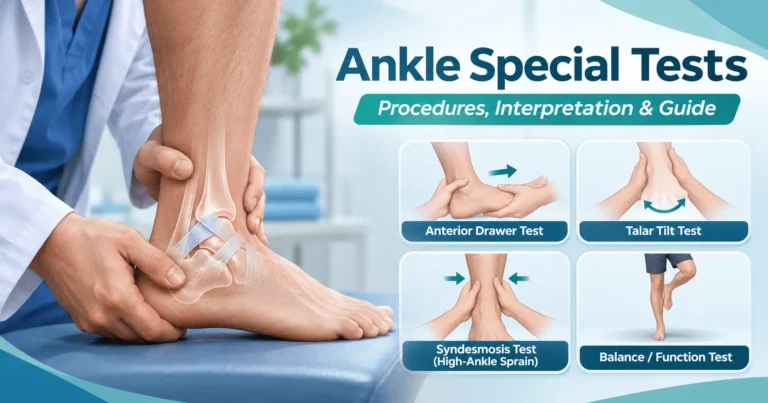
Ankle special tests help clinicians assess ligament injury, syndesmotic (high ankle) sprains, Achilles tendon rupture, and residual instability after injury. The most useful examination combines the injury mechanism, symptom location, palpation, range of motion, weight-bearing ability, and appropriately selected special tests. No single maneuver can confirm or exclude every ankle injury.
This evidence-informed guide is designed for physiotherapy students, physical therapists, athletic trainers, and other healthcare professionals. It explains what each test assesses, how to perform it, and how to interpret the findings. For the individual maneuvers, follow the linked detailed articles on Special Tests & Examination.
Quick reference: ankle special tests and what they assess
| Test | Primary clinical use | Finding that raises concern |
|---|---|---|
| Anterior drawer test | Anterior talofibular ligament (ATFL) injury | Excessive anterior talar movement or a soft end feel compared with the other ankle |
| Talar tilt test | Calcaneofibular ligament (CFL) injury with inversion stress | Increased inversion laxity or asymmetric end feel |
| Squeeze test | Syndesmotic (high ankle) injury | Reproduction of distal syndesmosis pain during proximal calf compression |
| External rotation stress test | Syndesmotic injury | Pain near the distal tibiofibular syndesmosis during stress |
| Deltoid ligament / eversion stress test | Medial ligament injury | Medial pain or abnormal laxity, interpreted cautiously |
| Thompson test | Achilles tendon rupture | Absent or markedly reduced plantarflexion when the calf is squeezed |
| Matles test | Achilles tendon rupture | Abnormal resting ankle position with the knee flexed |
| Functional assessment | Chronic ankle instability and recovery | Deficits in dorsiflexion, balance, reach, or hopping when appropriate |
Important: A positive test changes clinical suspicion; it is not the same as a definitive diagnosis. Many injuries coexist, and painful or swollen ankles may not tolerate reliable stress testing immediately after trauma.
Before special tests: screen the injured ankle
1. Take a focused history
Ask how the injury occurred, whether the ankle rolled inward or outward, whether the foot was planted during rotation, and whether a pop, immediate swelling, or difficulty walking followed. Establish the location of maximal pain, prior sprains, episodes of giving way, and any neurological symptoms.
2. Check for fracture risk and urgent concerns
Use the Ottawa Ankle Rules in the appropriate patient population to help decide whether radiographs are indicated. With pain in the malleolar zone, ankle radiographs are indicated when there is posterior-edge or tip tenderness of either malleolus, or inability to take four steps both immediately after injury and at assessment. With midfoot pain, assess navicular and fifth metatarsal base tenderness and the same weight-bearing criterion. The Ottawa rules address imaging decisions, not ligament severity.
Visible deformity, an open injury, a cold or poorly perfused foot, new major sensory loss, or other findings suggesting a serious injury require urgent medical assessment. Avoid provocative stress tests when fracture, dislocation, or severe acute injury is suspected.
3. Examine and compare sides
Inspect swelling and bruising; palpate relevant bony and soft-tissue structures; assess active movement and, when safe, passive movement. Compare the uninjured ankle’s laxity and resting position. Document pain, movement, apprehension, and the end feel rather than recording a test as positive or negative without explanation.
Lateral ankle ligament special tests
Anterior drawer test (ATFL)
The anterior drawer test assesses anterior translation of the talus relative to the tibia and is primarily used when an ATFL injury is suspected after an inversion mechanism.
- Position the patient sitting or lying with the lower leg supported and the ankle relaxed, typically in slight plantarflexion.
- Stabilize the distal tibia with one hand.
- With the other hand cupping the heel, gently draw the talus and foot forward while maintaining the tibial position.
- Compare excursion and end feel with the opposite ankle, noting whether the maneuver reproduces symptoms.
Interpretation: Increased anterior movement or a less firm end feel may support ATFL injury in the appropriate clinical context. Guarding, pain, anatomy, and examiner technique can affect the result. See the detailed ankle anterior drawer test guide.
Talar tilt test (CFL)
The inversion talar tilt test applies controlled varus stress to assess lateral ankle stability, especially the CFL. Support the distal lower leg, hold the hindfoot, and gently invert it while observing the range and end feel. Compare both sides and stop when significant pain or resistance occurs.
Interpretation: Clearly asymmetric inversion laxity supports a possible CFL injury, but a normal test does not reliably rule one out. Position and testing technique influence which structures are loaded. Read the talar tilt test article for further details.
When should lateral ligament tests be performed?
Immediate testing may be limited by pain, muscle guarding, and swelling. Research suggests that, when urgent injury has first been excluded, the accuracy of some ligament examinations can improve when repeated a few days later, often around days 2–7. This is a reassessment window, not a reason to delay necessary acute evaluation.
High ankle sprain: syndesmosis tests
A high ankle sprain affects the ligaments connecting the distal tibia and fibula. Suspect it after external rotation or dorsiflexion trauma, especially with pain above the ankle joint, tenderness over the anterior inferior tibiofibular ligament, and difficulty with push-off.
Squeeze test
With the patient relaxed, gently compress the tibia and fibula together at the mid-calf. The maneuver is considered suggestive of syndesmotic injury when it reproduces pain at the distal syndesmosis, rather than merely discomfort at the point of compression. Review the squeeze test guide.
External rotation stress test
With the lower leg stabilized and the knee flexed, the examiner carefully externally rotates the foot relative to the tibia, following a standardized clinical technique. Pain reproduced over the syndesmosis raises suspicion for high ankle injury. Avoid aggressive stress when severe injury is possible. See the external rotation test article.
Clinical interpretation: Neither test should be used alone. A systematic review of syndesmotic tests found no single maneuver with both high sensitivity and specificity; clinicians should combine the history, tenderness, functional findings, and imaging when warranted.
Medial ankle ligament tests
The deltoid ligament complex helps resist eversion and external rotation. Medial tenderness after an eversion or rotational mechanism may indicate deltoid injury, sometimes with associated fracture or syndesmotic injury. A gentle eversion stress examination may help assess medial stability, but marked pain, deformity, or suspected fracture warrants imaging assessment rather than repeated stress testing. See the deltoid ligament test.
Achilles tendon special tests
Thompson (calf squeeze) test
With the patient prone and feet hanging beyond the table, the clinician squeezes the calf and observes ankle plantarflexion. A lack of the expected plantarflexion response raises concern for Achilles tendon rupture. Compare the response with the opposite leg. A suspected rupture needs prompt clinical evaluation even if the patient can still move the foot. See the Thompson test article.
Matles test
In the Matles test, the patient lies prone with the knees flexed approximately 90 degrees. The examiner compares each ankle’s resting plantarflexion position. An ankle that falls into relatively greater dorsiflexion can support suspicion for an Achilles rupture. See the Matles test article. Both tests are interpreted alongside palpation, mechanism, and additional assessment as indicated.
Functional tests for chronic ankle instability
Ligament stress tests describe only part of the clinical picture. People with recurrent sprains or a feeling that the ankle “gives way” also need movement and function assessed.
- Weight-bearing lunge test: measures functional ankle dorsiflexion; document the method, distance or angle, and side-to-side difference.
- Single-leg stance: assesses static balance when the patient can safely weight bear; note time, errors, and testing conditions. Explore our single-leg balance stance test.
- Star Excursion Balance Test (SEBT): evaluates dynamic balance and reach control; measure and normalize reach distances when appropriate. Read the SEBT procedure and scoring guide.
- Hop or sport-specific tasks: consider only when symptoms, healing, and rehabilitation stage make the test safe.
The 2021 clinical practice guideline for lateral ankle sprains recommends repeated measurement of swelling, range of motion, laxity, static balance, and functional performance during rehabilitation.
A practical ankle examination sequence
- History: identify mechanism, onset, pain location, previous sprains, and function.
- Safety and fracture screen: apply imaging rules when appropriate and assess urgent neurovascular or structural concerns.
- Observation and palpation: record swelling, bruising, bony tenderness, and ligament or tendon tenderness.
- Motion and strength: check active motion and safe resisted movement.
- Targeted special tests: select lateral, syndesmotic, medial, or Achilles tests based on the suspected structure—not every test for every patient.
- Functional measures: add balance and movement tests when safe and relevant.
- Clinical synthesis: document the working diagnosis, differentials, need for imaging/referral, and reassessment plan.
Common mistakes to avoid
- Interpreting pain alone as proof of a ligament tear.
- Performing high-force stress tests on an acutely injured ankle before screening for fracture or serious injury.
- Ignoring the patient’s usual baseline laxity and not comparing the opposite side.
- Using one positive or negative special test as a definitive diagnosis.
- Failing to reassess when early swelling and guarding made the first examination inconclusive.
- Testing balance and hopping before the patient can safely tolerate weight bearing.
Frequently asked questions
What are the most common ankle special tests?
Frequently used tests include the anterior drawer, talar tilt, squeeze, external rotation stress, eversion stress, and Thompson test. Which tests matter most depends on the suspected injury and findings from the initial examination.
Which test assesses the ATFL?
The anterior drawer test is commonly used to assess the anterior talofibular ligament (ATFL). Palpation and related tests may add useful information; one test should not be interpreted in isolation.
Which tests help identify a high ankle sprain?
The squeeze and external rotation stress tests can support suspicion of syndesmotic injury. Neither independently confirms or rules it out, and further clinical assessment or imaging may be necessary.
Are ankle special tests accurate immediately after injury?
They can be harder to interpret during the first 48 hours because of swelling, guarding, and pain. Once serious injury is excluded, repeat examination after a few days may improve assessment of ligament laxity.
Are the Ottawa Ankle Rules an ankle ligament test?
No. The Ottawa Ankle Rules help clinicians decide when radiographs are needed after acute trauma; they do not identify which ligament is torn.
Key takeaway
Ankle special tests are most valuable when used selectively and in combination with history, palpation, functional examination, and appropriate imaging decisions. Anterior drawer and talar tilt inform lateral ligament assessment; squeeze and external rotation assess the syndesmosis; Thompson and Matles help examine the Achilles tendon. Document what each test actually showed rather than relying only on a positive or negative label.
References and further reading
- Martin RL, et al. Ankle Stability and Movement Coordination Impairments: Lateral Ankle Ligament Sprains Revision 2021. J Orthop Sports Phys Ther. 2021.
- Netterström-Wedin F, et al. Diagnostic Accuracy of Clinical Tests Assessing Ligamentous Injury of the Talocrural and Subtalar Joints. Systematic review and meta-analysis.
- Reliability and validity of physical examination tests for the assessment of ankle instability. Systematic review, 2022.
- Diagnostic accuracy of clinical tests assessing ligamentous injury of the ankle syndesmosis. Systematic review and meta-analysis, 2021.
- NSW Agency for Clinical Innovation. Ottawa ankle rules (adult), 2023.
- American Academy of Orthopaedic Surgeons. Achilles Tendon Rupture (Tear).