Trömner Sign: How to Perform the Test, Positive Results, Clinical Significance & Accuracy
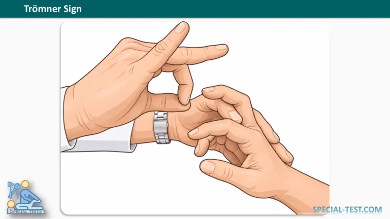
The Trömner sign (Tromner sign) is a neurological examination technique used to identify an exaggerated finger-flexor response that may indicate upper motor neuron dysfunction or cervical spinal cord compression. It is particularly relevant when assessing patients with suspected degenerative cervical myelopathy (DCM), a condition involving progressive dysfunction of the spinal cord in the neck.
The Trömner reflex is evaluated by stimulating the palmar surface of the middle finger and observing involuntary movements of the thumb and fingers. A positive response may provide evidence of corticospinal tract dysfunction, especially when accompanied by hyperreflexia, impaired hand coordination, weakness, or gait abnormalities.
Research suggests that the Trömner sign may be among the more sensitive neurological signs for detecting degenerative cervical myelopathy. However, a positive Trömner sign does not independently establish a diagnosis, and a negative result cannot reliably exclude spinal cord disease.
What Is the Trömner Sign?
The Trömner sign is a pathological or exaggerated finger-flexor reflex used during neurological examinations to assess possible abnormalities affecting descending motor pathways.
The test is commonly performed when a healthcare professional suspects an upper motor neuron lesion, particularly in patients experiencing symptoms associated with cervical spinal cord dysfunction.
The Trömner test is closely related to the Hoffmann sign. Both involve stimulation of the middle finger and observation of involuntary finger movements, but the stimulus is applied differently.
The primary purpose of the Trömner sign is to support the clinical assessment of possible corticospinal tract dysfunction, rather than to diagnose a specific neurological disorder.
Conditions in which an exaggerated finger-flexor response may be encountered include cervical myelopathy and other disorders affecting upper motor neuron pathways. The finding does not identify the precise location or cause of a lesion.
How to Perform the Trömner Sign Test
The Trömner test procedure is a simple bedside examination that generally requires no specialized equipment.
Patient Position
The patient should sit comfortably with the upper limb supported and the hand relaxed. The wrist and fingers should remain in a natural position, without active muscular contraction.
Examiner Position and Technique
- Position the hand: Support the patient’s hand while ensuring that the fingers remain relaxed.
- Stabilize the middle finger: Hold the patient’s middle finger lightly without restricting the movement being assessed.
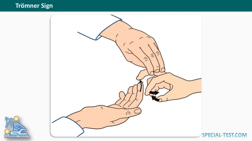
- Apply the stimulus: Deliver a quick, controlled upward flick or tap to the volar (palmar) surface of the distal phalanx of the middle finger.
- Observe the response: Look for involuntary flexion of the thumb and index finger, which may be accompanied by flexion of other fingers.
- Compare both hands: Repeat the maneuver on the opposite hand using a similar stimulus and observe any differences in response.
The stimulus should be brief and controlled rather than forceful. Painful stimulation is unnecessary and may interfere with accurate interpretation.
The examiner should document the presence, strength, symmetry, and reproducibility of the response.
See Also: Hoffmann’s Sign
What Is a Positive Trömner Sign?
A positive Trömner sign occurs when stimulation of the palmar surface of the distal middle finger produces involuntary flexion of the thumb and index finger.
This response may indicate increased excitability of finger-flexor reflex pathways associated with upper motor neuron dysfunction.
A positive finding becomes more clinically significant when other abnormalities are present, such as:
- Increased deep tendon reflexes (hyperreflexia).
- Impaired fine motor coordination of the hands.
- Muscle weakness or increased muscle tone.
- Difficulty walking or maintaining balance.
- Positive Hoffmann or Babinski signs.
- Other neurological findings suggesting spinal cord dysfunction.
However, some individuals without clinically significant spinal cord disease may demonstrate a finger-flexor response. Therefore, an isolated positive Trömner sign should not be interpreted as definitive evidence of cervical myelopathy.
What Does a Negative Trömner Sign Mean?
A negative Trömner sign indicates that the characteristic involuntary finger-flexion response is absent during testing.
Although a negative result may reduce clinical suspicion in certain circumstances, it does not rule out cervical myelopathy or other upper motor neuron disorders.
Patients with early or mild spinal cord dysfunction may have subtle findings, and examination results can vary depending on the technique, patient relaxation, and disease characteristics.
Progressive symptoms such as hand clumsiness, weakness, gait instability, or new bladder dysfunction require appropriate medical evaluation regardless of the Trömner test result.
Trömner Sign vs. Hoffmann Sign
The Trömner and Hoffmann signs are related neurological tests used to evaluate finger-flexor responses associated with possible corticospinal tract dysfunction.
Their main difference is the way the middle finger is stimulated.
| Feature | Trömner Sign | Hoffmann Sign |
|---|---|---|
| Stimulus location | Palmar surface of distal middle finger | Fingernail or distal middle finger |
| Technique | Quick upward flick or tap | Downward flick followed by sudden release |
| Positive response | Involuntary thumb and index finger flexion | Thumb flexion/adduction and possible index finger flexion |
| Clinical purpose | Assessment of possible upper motor neuron dysfunction | Assessment of possible upper motor neuron dysfunction |
| Diagnostic value | May support suspected cervical myelopathy | May support suspected cervical myelopathy |
Although both tests provide useful neurological information, neither should be used independently to confirm or exclude cervical spinal cord compression.
Diagnostic Accuracy of the Trömner Sign
The diagnostic performance of the Trömner sign has been examined in several clinical studies and systematic reviews.
A 2023 systematic review and meta-analysis published in Global Spine Journal identified the Trömner sign and hyperreflexia among the most sensitive clinical signs for degenerative cervical myelopathy. The review also identified the Trömner sign among tests demonstrating relatively high specificity in the included studies [1].
A 2025 scoping review published in Spinal Cord evaluated 16 studies and reported the following diagnostic accuracy ranges for the Trömner sign [2]:
- Sensitivity: 93–97%.
- Specificity: 79–100%.
These findings suggest that the test may be clinically useful in selected patient populations. However, the reported values should not be applied universally because study populations, examination methods, and reference standards differ.
An earlier diagnostic study involving 85 participants reported a sensitivity of 94% for the Trömner sign in cervical spondylotic myelopathy [3]. Nevertheless, positive responses were also common among participants with other cervical spine conditions, highlighting the risk of false-positive findings.
Another study examining physical signs of cervical cord compression reported a Trömner sign sensitivity of 72% and specificity of 79%. It also identified limitations in interobserver reliability, indicating that examination results may differ between clinicians [4].
Clinical interpretation: The available evidence supports including the Trömner sign in a comprehensive neurological examination, but no single accuracy estimate can reliably predict its performance in every clinical setting.
Clinical Importance in Cervical Myelopathy
Degenerative cervical myelopathy develops when age-related changes in the cervical spine contribute to compression and dysfunction of the spinal cord.
Patients may initially experience subtle neurological changes that are easily overlooked.
Common symptoms include difficulty fastening buttons, deterioration in handwriting, dropping objects, numbness or tingling in the hands, impaired balance, and changes in walking ability.
During examination, clinicians may identify increased reflexes, abnormal finger-flexor responses, impaired coordination, spasticity, or pathological plantar responses.
The Trömner sign can contribute to recognition of these neurological abnormalities, particularly when the patient’s symptoms suggest cervical spinal cord involvement.
However, it is essential to distinguish cervical myelopathy from conditions such as cervical radiculopathy, peripheral neuropathy, and other neurological disorders.
When cervical myelopathy is suspected, magnetic resonance imaging (MRI) of the cervical spine is commonly used to evaluate spinal cord compression and associated structural abnormalities.
Limitations of the Trömner Test
Despite its clinical usefulness, the Trömner sign has several limitations.
Examiner technique: Differences in stimulus strength, direction, and finger positioning may influence the response.
Patient-related factors: Anxiety, muscular tension, discomfort, and the ability to relax may affect reflex assessment.
False-positive results: An exaggerated finger-flexor response can occur without clinically significant cervical spinal cord disease.
False-negative results: Some patients with confirmed myelopathy do not demonstrate the sign.
Limited anatomical localization: A positive result does not identify the exact spinal level or underlying cause of neurological dysfunction.
Variable reliability: Research has demonstrated differences in agreement between examiners, emphasizing the importance of standardized examination techniques [4].
For these reasons, the Trömner sign should be interpreted alongside the patient’s symptoms, neurological examination, and appropriate investigations.
When Is Further Medical Evaluation Necessary?
Patients with a positive Trömner sign and symptoms suggesting progressive cervical myelopathy should undergo appropriate clinical assessment.
Particular attention should be given to progressive hand weakness, deterioration in fine motor coordination, increasing gait instability, frequent falls, or new sensory disturbances.
Rapidly worsening weakness, substantial deterioration in walking ability, or new bowel or bladder dysfunction associated with neurological symptoms warrants urgent medical evaluation.
Clinical decisions should be guided by the complete neurological presentation rather than repetition of an isolated reflex test.
Frequently Asked Questions (FAQ)
1. What is the Trömner sign used for?
The Trömner sign is used to assess an involuntary finger-flexor response that may indicate upper motor neuron dysfunction. It is particularly useful as part of the neurological examination of patients with suspected cervical myelopathy.
2. How do you perform the Trömner test?
The examiner supports the relaxed hand and delivers a brief upward flick to the palmar surface of the distal middle finger. Involuntary flexion of the thumb and index finger is observed.
3. What does a positive Trömner sign indicate?
A positive Trömner sign may suggest increased reflex excitability associated with corticospinal tract dysfunction. It becomes more meaningful when accompanied by other findings consistent with cervical spinal cord disease.
4. Is the Trömner sign the same as the Hoffmann sign?
No. Both tests examine finger-flexor responses, but the Trömner sign involves stimulation of the palmar surface of the distal middle finger, whereas the Hoffmann sign typically involves flicking the distal finger or fingernail and allowing it to rebound.
5. How accurate is the Trömner sign?
A 2025 scoping review reported sensitivity ranging from 93% to 97% and specificity ranging from 79% to 100% in the studies it summarized. Other research has reported lower values, so accuracy depends on the patient population and examination methods [2,4].
6. Can a healthy person have a positive Trömner sign?
Yes. A finger-flexor response may occur in individuals without clinically significant spinal cord disease. An isolated positive finding is insufficient to diagnose an upper motor neuron lesion.
7. Does a negative Trömner sign rule out cervical myelopathy?
No. A negative result cannot independently exclude cervical myelopathy. Persistent or progressive neurological symptoms require further evaluation regardless of the test result.
8. Is the Trömner sign associated with cervical spinal cord compression?
Yes. An exaggerated Trömner response may be observed in patients with cervical spinal cord compression, including degenerative cervical myelopathy. However, the test does not establish the presence or severity of compression by itself.
9. Can the Trömner sign determine the severity of myelopathy?
Not reliably through routine bedside examination alone. Some research has investigated quantitative measurement of the Trömner response and its association with cervical cord compression, but the standard clinical test is not a validated stand-alone severity measure [5].
10. What should be done after a positive Trömner test?
The clinician should assess the patient’s neurological symptoms and perform a comprehensive examination. If cervical myelopathy is suspected, further evaluation, commonly including cervical spine MRI, may be indicated.
Conclusion
The Trömner sign is an important neurological examination finding that can help clinicians identify possible upper motor neuron dysfunction and cervical spinal cord involvement.
It is performed by applying a quick stimulus to the palmar surface of the distal middle finger and observing involuntary thumb and finger flexion.
Clinical research suggests that the Trömner sign can demonstrate relatively high sensitivity for degenerative cervical myelopathy in selected populations. Nevertheless, diagnostic accuracy varies between studies, and a positive response does not confirm spinal cord disease.
For reliable clinical assessment, the Trömner test should always be interpreted together with patient symptoms, muscle strength, coordination, tendon reflexes, gait assessment, and appropriate diagnostic imaging.
References
- Jiang Z, et al. The Value of Clinical Signs in the Diagnosis of Degenerative Cervical Myelopathy: A Systematic Review and Meta-analysis. Global Spine Journal. 2023. PubMed – PMID: 37903098
- Sharma S, et al. Clinical Signs and Symptoms for Degenerative Cervical Myelopathy: A Scoping Review of Case-Control Studies to Facilitate Early Diagnosis Among Healthcare Professionals With Stakeholder Engagement. Spinal Cord. 2025;63(3):171–180. PubMed – PMID: 40011743
- The Significance of the Trömner Sign in Cervical Spondylotic Myelopathy Patient. PubMed – PMID: 27404855
- Kato Y, et al. Accuracy and Reliability of Physical Signs as a Diagnostic Tool for Cervical Cord Compression: A Cross-Sectional Study. Spine Surgery and Related Research. 2024. PubMed – PMID: 40223840
- Chang CW, Chang KY, Lin SM. Quantification of the Trömner Signs: A Sensitive Marker for Cervical Spondylotic Myelopathy. European Spine Journal. 2011;20(6):923–927. PubMed – PMID: 21221662
- Hoffman Sign: A Neurological Test for Upper Motor Neuron Lesions. Orthofixar. Read on Orthofixar
- Cervical Myelopathy: Warning Signs, Diagnosis & Treatment. Orthofixar. Read on Orthofixar