Wada Test: Purpose, Procedure, Interpretation, and Risks
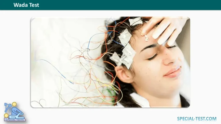
The Wada test, also known as the intracarotid amobarbital procedure (IAP) or intracarotid sodium amobarbital test, is a specialized neurological examination used to determine which hemisphere of the brain is responsible for essential cognitive functions, particularly language and memory.
The test is primarily performed during the presurgical evaluation of patients with drug-resistant epilepsy, especially those being considered for temporal lobe surgery. In selected cases, it may also be used before brain tumor surgery or other neurosurgical procedures involving regions associated with language and memory.
By temporarily suppressing the function of one cerebral hemisphere, the Wada test allows neurologists to evaluate the functional capabilities of the opposite hemisphere. This information helps surgeons estimate the risk of postoperative speech difficulties, memory impairment, or severe amnesia.
Although functional magnetic resonance imaging (fMRI) has increasingly replaced the Wada test in routine presurgical evaluations, the procedure remains valuable in selected complex cases.
What Is the Purpose of the Wada Test?
The primary purpose of the Wada test is to establish cerebral language dominance and assess memory function in each hemisphere before brain surgery.
Its principal clinical applications include:
- Language lateralization: Determining whether the left hemisphere, right hemisphere, or both hemispheres contribute significantly to language function.
- Memory assessment: Evaluating the ability of each hemisphere to support memory when the opposite hemisphere is temporarily suppressed.
- Epilepsy surgery planning: Assessing the risk of postoperative cognitive deficits, particularly before temporal lobectomy.
- Brain tumor surgery: Providing additional information about language and memory representation when surgery may affect critical functional brain regions.
- Risk assessment for postoperative amnesia: Identifying patients who may have insufficient memory function in the hemisphere that will remain unaffected by surgery.
The test is particularly important when conventional neuropsychological examinations and neuroimaging studies provide conflicting or inconclusive results.
How Is the Wada Test Performed?
The Wada test is an invasive diagnostic procedure performed by a multidisciplinary medical team, typically including an interventional neuroradiologist, neurologist, neuropsychologist, and specialized nursing staff.
The patient remains awake and able to communicate throughout the neurological testing.
1. Patient Preparation
Before the procedure, the medical team reviews the patient’s neurological history, medications, vascular anatomy, and relevant imaging studies.
Baseline language and memory assessments may be performed to establish the patient’s normal cognitive performance.
An intravenous line is placed, and appropriate physiological monitoring is established.
2. Catheter Insertion and Cerebral Angiography
An interventional neuroradiologist inserts a catheter into an artery, usually through the femoral artery in the groin, and advances it toward one of the internal carotid arteries under imaging guidance.
Cerebral angiography is performed to evaluate the vascular anatomy and identify potential cross-flow between cerebral hemispheres.
This assessment is important because unusual vascular connections can influence the distribution of the anesthetic medication and affect interpretation of the results.
3. Administration of Sodium Amobarbital
A short-acting anesthetic, traditionally sodium amobarbital (Amytal), is injected through the catheter into one internal carotid artery.
Alternative medications, including methohexital or etomidate, may be used according to institutional protocols.
The medication temporarily suppresses neuronal activity in the cerebral regions supplied by the injected artery.
For example:
- Injection into the left internal carotid artery primarily suppresses function in the left cerebral hemisphere.
- Injection into the right internal carotid artery primarily suppresses function in the right cerebral hemisphere.
This reversible suppression allows clinicians to assess how the opposite hemisphere performs essential cognitive tasks.
4. EEG and Neurological Monitoring
Electroencephalography (EEG) is commonly used throughout the procedure to monitor changes in cerebral electrical activity.
The injected anesthetic typically produces temporary slowing of EEG activity in the affected hemisphere.
The neurologist also evaluates motor function, as transient weakness of the arm and leg opposite the injected hemisphere can help confirm the medication’s neurological effect.
EEG findings and motor examination help determine when language and memory assessments can be performed reliably.
5. Language Assessment
During the anesthetic effect, the patient undergoes several language-related tasks.
These may include:
- Naming familiar objects or pictures.
- Counting aloud.
- Reading words or sentences.
- Repeating spoken phrases.
- Following verbal instructions.
- Answering simple questions.
Significant language disturbance following injection suggests that the suppressed hemisphere makes an important contribution to language function.
For example, temporary aphasia after left-sided injection supports left-hemisphere language dominance, although the complete test results must be considered.
6. Memory Assessment
Memory testing evaluates whether the nonsuppressed hemisphere can support the encoding and subsequent recognition or recall of information.
During the period of temporary hemispheric suppression, the patient is shown pictures, objects, words, or other materials.
After the anesthetic effect has dissipated, the examiner asks the patient to recognize or recall the previously presented information.
Performance is assessed according to the institution’s standardized protocol.
7. Testing the Opposite Hemisphere
Once the patient has recovered sufficiently and the medication’s neurological effects have resolved, the procedure may be repeated on the opposite side.
Testing each hemisphere separately allows clinicians to compare language performance and memory capacity.
How Are Wada Test Results Interpreted?
Wada test interpretation focuses on the distribution of language function and the relative memory capabilities of the two cerebral hemispheres.
Language Dominance
The results may demonstrate:
- Left-hemisphere dominance: Language function depends predominantly on the left hemisphere.
- Right-hemisphere dominance: Language function depends predominantly on the right hemisphere.
- Bilateral language representation: Both hemispheres make meaningful contributions to language.
The results help surgeons understand the potential consequences of operating on the language-dominant hemisphere.
Importantly, the Wada test primarily evaluates hemispheric dominance rather than precisely identifying individual language areas within the brain.
Memory Function
Memory assessment focuses on whether the hemisphere that will remain intact after surgery has sufficient functional reserve.
For example, in a patient undergoing left temporal lobe surgery, clinicians are particularly concerned about the memory capabilities of the right hemisphere.
Poor memory performance when the hemisphere planned for surgery is temporarily suppressed may indicate an increased risk of significant postoperative memory impairment.
However, Wada memory results are not perfectly predictive of postoperative outcomes and should be interpreted alongside neuropsychological testing, MRI findings, seizure localization, and other clinical data.
Risks and Complications of the Wada Test
Although the Wada test is generally performed safely in experienced medical centers, it involves arterial catheterization and administration of an anesthetic directly into the cerebral circulation.
Potential complications include:
- Bleeding or hematoma at the catheter insertion site.
- Temporary confusion or altered consciousness.
- Seizures.
- Allergic reactions to angiographic contrast material.
- Carotid artery injury or dissection.
- Transient ischemic attack (TIA).
- Stroke, which is uncommon but potentially serious.
A retrospective study of 677 patients reported complications in 10.9% of procedures, including transient neurological effects and less frequent vascular complications. Reported complication rates vary substantially depending on how complications are defined and the populations studied.
The potential risks must be considered against the clinical value of the information obtained.
Wada Test vs. Functional MRI (fMRI)
Functional MRI has become an important noninvasive alternative to Wada testing during presurgical epilepsy assessment.
| Feature | Wada Test | Functional MRI |
|---|---|---|
| Invasiveness | Invasive | Noninvasive |
| Main technique | Temporary pharmacological suppression | Brain activity mapping using blood-oxygenation changes |
| Language assessment | Hemispheric functional testing | Language activation and lateralization |
| Memory assessment | Direct testing during temporary hemispheric suppression | Task-based assessment and prediction of memory outcomes |
| Main limitations | Vascular risks and variable memory prediction | Task dependence and potentially inconclusive results |
| Current role | Selected complex cases | Frequently used in routine presurgical evaluation |
According to the American Academy of Neurology practice guideline, fMRI may be considered as an alternative to the intracarotid amobarbital procedure for language lateralization in several epilepsy populations.
However, the Wada test may remain useful when fMRI results are inconclusive, language dominance is atypical, or there is particular concern about severe postoperative amnesia.
Frequently Asked Questions (FAQs)
1. What does the Wada test measure?
The Wada test evaluates the contribution of each cerebral hemisphere to language and memory. It helps determine language dominance and assess the potential risk of cognitive impairment following certain brain surgeries.
2. Is the Wada test painful?
The procedure is generally performed using local anesthesia at the catheter insertion site. Patients may experience temporary discomfort during catheter placement or other parts of the examination. The medical team monitors the patient throughout the procedure.
3. Is the patient awake during the Wada test?
Yes. Patients are kept awake and sufficiently alert to complete language and memory tasks. The purpose is to assess cognitive performance while one cerebral hemisphere is temporarily affected by an anesthetic.
4. How long do the effects of the Wada test last?
The neurological effects of the injected medication usually last only several minutes, although the exact duration depends on the anesthetic and dose used. The overall appointment takes longer because of preparation, angiography, testing, and postprocedural observation.
5. Why is the Wada test performed before epilepsy surgery?
The test helps identify which hemisphere supports language and whether the opposite hemisphere has adequate memory capacity. This information can help surgeons estimate the risk of postoperative aphasia or memory impairment.
6. Can the Wada test cause permanent brain damage?
Permanent neurological complications are uncommon, but serious vascular events, including stroke, are possible. Patients should discuss their individual risk factors with the treating neurological team.
7. Can functional MRI replace the Wada test?
Functional MRI can replace Wada testing in many presurgical evaluations, particularly for language lateralization. However, the Wada test may still be indicated when noninvasive findings are unclear or when there is a substantial concern about postoperative global amnesia.
8. Is the Wada test still commonly performed?
Its use has decreased as noninvasive brain-mapping methods have improved. It is no longer routinely required for every epilepsy surgery candidate, but it remains an option for selected patients with complex or uncertain findings.
Conclusion
The Wada test is an important diagnostic tool for assessing cerebral language dominance and memory function in selected patients undergoing evaluation for epilepsy surgery or other neurosurgical procedures.
Through temporary suppression of one cerebral hemisphere, the test provides information about the functional capabilities of the opposite hemisphere and helps estimate the likelihood of postoperative neurological deficits.
Despite its historical importance, the Wada test has limitations, particularly regarding the prediction of specific memory outcomes, and carries risks associated with invasive cerebral angiography.
Modern techniques such as functional MRI and comprehensive neuropsychological assessment have reduced the need for routine Wada testing. Nevertheless, it remains clinically relevant when noninvasive investigations are inconclusive or when a patient may be at increased risk of severe postoperative memory impairment.
References
- Trenerry MR, Loring DW. Intracarotid amobarbital procedure: The Wada test. Neuroimaging Clinics of North America. 1995;5(4):721–728. PubMed – PMID: 8564293
- Sharan A, Ooi YC, Langfitt J, Sperling MR. Intracarotid amobarbital procedure for epilepsy surgery. Epilepsy & Behavior. 2011;20(2):209–213. PubMed – PMID: 21190900
- Szaflarski JP, et al. Practice guideline summary: Use of fMRI in the presurgical evaluation of patients with epilepsy. Neurology. 2017. PubMed – PMID: 28077494 | NCBI Full Text
- Schmid E, et al. Diagnostic accuracy of functional magnetic resonance imaging, Wada test, magnetoencephalography, and functional transcranial Doppler sonography for memory and language outcome after epilepsy surgery: A systematic review. Epilepsia. 2018;59(12):2305–2317. PubMed – PMID: 30374948
- Roles of fMRI and Wada tests in the presurgical evaluation of language functions in temporal lobe epilepsy. 2022. NCBI – Full-Text Review
- Complications during the Wada test. Epilepsy & Behavior. 2008. PubMed – PMID: 18590981
- Beyond the Wada: An updated approach to pre-surgical language and memory testing. Epilepsy Research. 2021. PubMed – PMID: 34082393
- Quantitative EEG is an objective, sensitive and reliable indicator of transient anesthetic effects during Wada tests. PubMed – PMID: 25580802 | NCBI Full Text