Hoover’s Test (Hoover’s Sign): Procedure, Positive Results, Interpretation & Accuracy
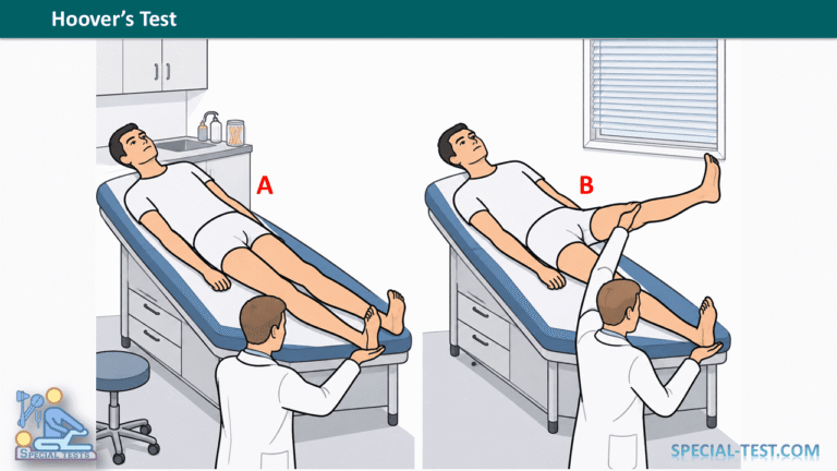
Hoover’s test, also known as Hoover’s sign, is a neurological examination technique used to differentiate functional leg weakness from weakness caused by structural neurological disease. It evaluates the relationship between voluntary hip extension and involuntary hip extension produced during movement of the opposite leg.
First described by Dr. Charles Franklin Hoover in 1908, this bedside examination remains an important clinical tool for assessing patients with unexplained unilateral lower limb weakness. It is particularly useful when evaluating suspected functional neurological disorder (FND), previously called conversion disorder or functional neurological symptom disorder.
Hoover’s sign is based on the principle that voluntary hip flexion normally produces an automatic extension force in the opposite lower limb. In patients with functional weakness, voluntary hip extension may appear markedly reduced, while automatic extension strength remains relatively preserved.
A positive Hoover’s sign supports the diagnosis of functional limb weakness. However, the test must be interpreted alongside the patient’s medical history, neurological examination, and other clinical findings. [1,2]
What Is Hoover’s Test Used For?
The primary purpose of Hoover’s test is to identify inconsistencies between voluntary and automatic muscle activation in patients presenting with lower limb weakness.
Clinical indications include:
- Functional leg weakness: Identifying positive signs of functional neurological disorder affecting one lower extremity.
- Differential diagnosis of leg paresis: Helping distinguish functional weakness from weakness associated with structural neurological lesions.
- Neurological examination: Assessing patients with unexplained unilateral weakness or suspected functional hemiparesis.
- Stroke assessment: Providing supportive clinical information when evaluating stroke-like presentations, without replacing urgent stroke investigations.
- Assessment of motor function: Comparing voluntary hip extension with automatic muscle activation during contralateral hip flexion.
The Hoover test is most useful in patients with unilateral leg weakness and adequate strength in the opposite leg, allowing meaningful comparison between both lower limbs.
Importantly, functional weakness is a genuine neurological symptom. A positive Hoover’s sign does not demonstrate malingering, deliberate symptom production, or lack of cooperation. [1,3]
How to Perform Hoover’s Test
Hoover’s test is performed with the patient lying supine on an examination table. The examiner assesses the downward pressure generated by each heel during voluntary and automatic movements.
Step 1: Patient Position
Ask the patient to lie comfortably in the supine position, with both lower limbs extended and relaxed.
The examiner stands at the foot of the examination table and places one hand beneath each heel or ankle.
Explain the examination and ensure that the patient understands the instructions.
Step 2: Assess Baseline Hip Extension Strength
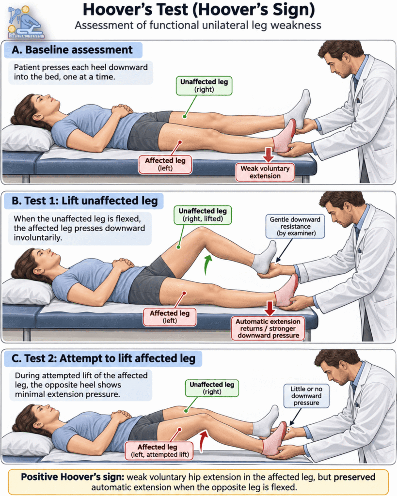
Ask the patient to press one heel downward against the examiner’s hand, as though pushing the leg into the examination table.
Repeat this movement with the opposite leg.
Compare the downward pressure generated by each limb.
In patients with functional leg weakness, the affected limb may demonstrate reduced voluntary hip extension strength.
Step 3: Assess Automatic Extension of the Affected Leg
Maintain the examiner’s hand beneath the heel of the affected limb.
Ask the patient to lift the unaffected leg off the examination table by flexing the hip. Gentle downward resistance may be applied to the lifting leg.
During this movement, observe the pressure exerted by the affected heel against the examiner’s hand.
Normally, hip flexion on one side produces automatic hip extension on the opposite side.
In functional weakness, the affected leg may generate a substantially stronger downward force during this automatic movement than during direct voluntary hip extension.
Step 4: Assess Contralateral Extension During Affected-Leg Flexion
Place the examiner’s hand beneath the heel of the unaffected limb.
Ask the patient to attempt to raise the affected leg while keeping the opposite leg on the table.
Observe the downward pressure beneath the unaffected heel.
In patients with functional leg weakness, the expected automatic extension pressure beneath the unaffected leg may be reduced or absent during the attempted movement.
Compare these findings with the baseline examination and the previous maneuver.
Step 5: Compare the Findings
Assess whether there is a significant difference between voluntary and automatic hip extension.
The central feature of Hoover’s sign is inconsistency between reduced voluntary strength and preserved automatic strength in the affected lower limb. [1,4]
Hoover’s Test: Positive and Negative Results
Positive Hoover’s Sign
A positive Hoover’s sign is present when the affected leg demonstrates weak voluntary hip extension but produces noticeably stronger, potentially normal, automatic hip extension when the opposite hip is flexed.
A second supportive finding is reduced or absent downward pressure beneath the unaffected heel when the patient attempts to lift the affected leg.
The appearance of preserved strength during automatic movement suggests functional leg weakness rather than a fixed loss of motor power caused solely by structural neurological damage.
Negative Hoover’s Sign
A negative Hoover’s sign occurs when the characteristic inconsistency between voluntary and automatic hip extension is not demonstrated.
For example, an affected leg may remain weak during both voluntary and automatic movements.
However, a negative result does not establish an organic neurological cause or exclude functional neurological disorder.
Hoover’s Test Interpretation
| Examination finding | Clinical interpretation |
|---|---|
| Weak voluntary hip extension with stronger automatic extension of the affected leg | Positive Hoover’s sign; supports functional weakness |
| Minimal pressure beneath the unaffected heel during attempted elevation of the affected leg | Additional finding supporting functional weakness |
| Weakness during both voluntary and automatic hip extension | No characteristic positive sign demonstrated |
| Unclear or inconsistent results affected by pain or examination difficulties | Indeterminate; further assessment required |
Hoover’s Test Sensitivity and Specificity
The diagnostic accuracy of Hoover’s sign has been examined in clinical research.
A prospective cohort study published in the Journal of Psychosomatic Research evaluated Hoover’s sign among patients presenting with suspected stroke.
The study included 337 patients, of whom 124 had leg weakness. Eight patients with leg weakness received a diagnosis of functional disorder.
The researchers reported:
- Sensitivity: 63%
- Specificity: 100%
These findings indicate that a positive Hoover’s sign was highly specific for functional weakness in this study population, although the examination did not identify every patient with functional weakness.
The results should be interpreted cautiously because relatively few patients in the cohort had functional weakness.
A 2025 review of validated functional motor weakness examinations also emphasized that Hoover’s sign should not be used as an isolated diagnostic test. [1,2]
Limitations of Hoover’s Sign
Although Hoover’s test is clinically valuable, several factors can affect its interpretation.
Important limitations include:
- Pain-related weakness: Hip, back, or lower limb pain may interfere with the patient’s ability to generate force.
- Bilateral leg weakness: Assessment is less reliable when both lower extremities are weak because an adequately strong comparison limb is needed.
- Neurological conditions: Certain neurological deficits, including hemineglect or apraxia, may complicate interpretation.
- Patient positioning and technique: Inconsistent examiner hand placement, inadequate instructions, or variations in resistance can affect results.
- Coexisting conditions: Functional weakness and structural neurological disease may occur in the same patient.
The test should form part of a comprehensive neurological assessment, including muscle strength, tone, deep tendon reflexes, sensation, coordination, and gait examination when appropriate.
Sudden-onset leg weakness, particularly when accompanied by facial weakness, speech disturbance, or other acute neurological symptoms, requires emergency evaluation for stroke or other serious neurological disease.
Clinical Significance of Hoover’s Test
Hoover’s sign is one of the best-studied positive physical examination findings associated with functional limb weakness.
Unlike an approach based entirely on excluding structural neurological disease, demonstrating a positive Hoover’s sign provides supportive evidence of altered voluntary motor control with preservation of automatic motor function.
This distinction is important because functional neurological disorder is a recognized clinical condition that can cause substantial physical disability.
When appropriately explained, the examination may also help patients understand how movement and strength can change under different circumstances.
A positive sign can therefore contribute to diagnosis, patient education, and planning appropriate rehabilitation, including specialist physiotherapy when indicated. [3,5]
Frequently Asked Questions (FAQs)
1. What is Hoover’s test?
Hoover’s test is a neurological bedside examination used to identify functional leg weakness. It compares voluntary hip extension strength with the automatic hip extension produced when the opposite hip is flexed.
2. What does a positive Hoover’s sign indicate?
A positive Hoover’s sign indicates that voluntary hip extension appears weak, while automatic hip extension is relatively preserved. This pattern supports functional neurological weakness but does not establish the diagnosis on its own.
3. How do you perform Hoover’s test?
The patient lies supine while the examiner places their hands beneath the heels. The patient is asked to press each heel downward and then raise each leg separately. The examiner compares the downward forces generated during voluntary and automatic movements.
4. What is the sensitivity and specificity of Hoover’s test?
In one prospective study involving patients with suspected stroke and leg weakness, Hoover’s sign demonstrated 63% sensitivity and 100% specificity for functional weakness. These estimates are based on a small functional weakness subgroup and should not be assumed to apply to every clinical setting.
5. Does a positive Hoover’s sign mean the patient is faking weakness?
No. A positive Hoover’s sign does not establish intentional symptom production or malingering. Functional neurological weakness is a genuine condition involving altered motor control.
6. Can Hoover’s sign distinguish functional weakness from stroke?
Hoover’s sign can support the differential diagnosis of functional weakness in patients presenting with stroke-like symptoms. However, it cannot independently exclude stroke, and urgent neurological assessment remains essential when stroke is suspected.
7. Is Hoover’s test reliable in bilateral leg weakness?
Hoover’s sign is most useful in unilateral leg weakness. When both legs are weak, the absence of a strong comparison limb limits the reliability of the examination.
Conclusion
Hoover’s test is a valuable neurological examination technique for assessing functional lower limb weakness. Its primary diagnostic feature is the discrepancy between reduced voluntary hip extension and preserved automatic hip extension during contralateral hip flexion.
A positive Hoover’s sign provides evidence supporting functional neurological weakness, particularly when accompanied by other consistent examination findings.
Despite its high reported specificity in selected clinical studies, the test should never replace a comprehensive neurological assessment or appropriate investigations.
References
- Dolbow J, et al. (2025). A practical guide to assessing functional motor weakness: a review of validated techniques. Journal of Neurology. NCBI – PubMed Central
- McWhirter L, Stone J, Sandercock P, Whiteley W. (2011). Hoover’s sign for the diagnosis of functional weakness: a prospective unblinded cohort study in patients with suspected stroke. Journal of Psychosomatic Research, 71(6), 384–386. PubMed – PMID: 22118379
- Stone J, et al. (2021). A practical review of functional neurological disorder (FND) for the general physician. Clinical Medicine. NCBI – PubMed Central
- Mehndiratta MM, Kumar M, Nayak R, Garg H, Pandey S. (2014). Hoover’s sign: Clinical relevance in Neurology. Journal of Postgraduate Medicine, 60(3), 297–299. PubMed – PMID: 25121372
- Nielsen G, et al. (2015). Physiotherapy for functional motor disorders: a consensus recommendation. Journal of Neurology, Neurosurgery & Psychiatry, 86(10), 1113–1119. NCBI – PubMed Central
- Orthofixar. Hoover Test: Differentiate Organic vs Functional Leg Weakness. Orthofixar – Hoover Test