Hip Examination: History Taking and Clinical Examination
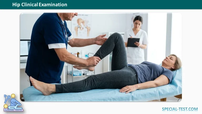
The hip joint is a deep ball-and-socket synovial joint that plays a vital role in weight-bearing and mobility. Because pain arising from the hip may be referred to the groin, thigh, knee, or buttock, a systematic clinical examination is essential for identifying the underlying pathology.
A complete hip examination includes:
- History taking
- Inspection
- Palpation
- Assessment of range of motion
- Special tests
- Neurovascular examination
- Examination of adjacent joints (lumbar spine, pelvis, knees, ankles, and feet)
History Taking
A thorough history often provides the diagnosis before the physical examination begins.
Pain
Pain is the most common presenting complaint. Important aspects include:
- Onset
- Acute pain suggests trauma, septic arthritis, reactive arthritis, muscle injury, or fracture.
- Insidious onset suggests osteoarthritis, inflammatory arthritis, osteonecrosis, tuberculosis, or tumors.
- Duration
- Sudden onset usually indicates trauma or infection.
- Chronic pain favors degenerative or inflammatory conditions.
- Character
- Sharp, stabbing pain is common after trauma.
- Dull aching pain occurs in early osteoarthritis or avascular necrosis.
- Throbbing pain is often associated with infection.
- Diurnal variation
- Morning stiffness suggests inflammatory arthritis such as rheumatoid arthritis or ankylosing spondylitis.
- Night pain may occur with infection, tumors, or tuberculosis.
- Pain during the first few steps after resting is typical of arthritis.
- Pain appearing after prolonged activity is more suggestive of early osteonecrosis or overuse conditions.
- Progression
- Gradually worsening pain suggests degenerative disease.
- Sudden worsening after a chronic course may indicate femoral head collapse in osteonecrosis or a pathological fracture.
Pain Location
The site of pain provides useful diagnostic clues:
| Pain Location | Possible Cause |
|---|---|
| Groin | Intra-articular hip pathology |
| Buttock | Sacroiliac joint or lumbar spine disease |
| Posterior thigh | Lumbar radiculopathy |
| Lateral hip | Greater trochanteric pain syndrome |
| “C-sign” (patient cups the lateral hip with thumb and fingers) | Femoroacetabular impingement (FAI) |
| Anterior knee (especially unexplained) | Referred pain from the hip |
Patients with persistent knee pain should always undergo a hip examination because hip disorders commonly present with referred knee pain.
Limp
Limp is one of the most reliable indicators of hip pathology.
Determine:
- Time of onset
- Relation to pain
- Progression
- Need for walking aids
Important considerations include:
- Tuberculosis of the hip often presents with limp before pain.
- Painless limp suggests:
- Developmental dysplasia of the hip (DDH)
- Coxa vara
- Cerebral palsy
- Poliomyelitis
- Healed hip infection with deformity
- Ankylosis
Stiffness
Ask about:
- Difficulty getting out of bed
- Difficulty putting on shoes or socks
- Morning stiffness
- Duration of stiffness
Stiffness may result from:
- Muscle spasm
- Synovitis
- Inflammatory arthritis
- Osteoarthritis
- Prolonged immobilization
Morning stiffness lasting more than 30–60 minutes strongly suggests inflammatory arthritis.
Deformity
Ask whether the patient has noticed:
- Limb shortening
- Rotational deformity
- Flexion deformity
- Progressive deformity
Patients usually recognize limb shortening more readily than rotational deformities.
Swelling
Because the hip joint lies deep beneath muscles, swelling is uncommon.
Ask about:
- Progressive swelling
- Groin masses
- Gluteal swelling
- Previous abscesses
- Sinus formation
Large swellings may occur with:
- Tuberculosis
- Septic arthritis
- Soft tissue tumors
- Iliopsoas abscess
Trauma History
If trauma occurred, document:
- Mechanism of injury
- Road traffic accident
- Fall from standing height
- Fall from height
- Sports injury
- Crush injury
- Slip or twisting injury
- Site of impact
- Ability to walk after injury
- Ability to bear weight
- Associated injuries
- Initial treatment received
Functional Disability
Assess current functional limitations, including:
- Walking distance
- Stair climbing
- Sitting cross-legged
- Squatting
- Getting into a car
- Wearing shoes and socks
- Need for walking aids
Also determine the patient’s pre-injury mobility.
Important Negative History
Always ask about:
- Fever
- Weight loss
- Night sweats
- Tuberculosis exposure
- Pain in other joints
- Back pain
- Pregnancy
- Previous tuberculosis treatment
- Symptoms in the opposite hip
- Ipsilateral and contralateral knee
- Ankle and foot symptoms
Past Medical History
Include:
- Previous tuberculosis
- Previous hip trauma
- Hip surgery
- Congenital hip disorders
- Developmental dysplasia
- Osteoporosis
- Diabetes mellitus
- Hypertension
- Renal disease
- Liver disease
- Respiratory disease
- Neurological disorders
- Connective tissue disorders
- Hematological disorders
- Organ transplantation
Also assess fitness for anesthesia if surgery is anticipated.
Personal History
Ask about:
- Occupation
- Activity level
- Sports participation
- Diet
- Smoking
- Alcohol consumption
- Recreational drug use
Family History
Ask about:
- Developmental dysplasia of the hip
- Inflammatory arthropathies
- Storage disorders
- Hereditary skeletal dysplasias
Inspection
Observe the patient while standing, walking, and lying supine.
Inspection from the Front
Assess:
- Overall posture
- Pelvic alignment
- Limb position
- Hip flexion deformity
- External or internal rotation
- Knee alignment
- Foot position
- Pelvic tilt
- ASIS symmetry
- Muscle wasting (especially quadriceps)
- Adductor muscle prominence or spasm
- Hernias
- Perineal widening
Inspect the skin for:
- Swelling
- Surgical scars
- Sinuses
- Loss of skin creases
- Dilated veins
- Discoloration
Inspection from the Side
Look for:
- Lumbar lordosis
- Pelvic tilt
- Hip flexion deformity
- Knee flexion
- Equinus deformity at the ankle
- Trochanteric prominence
Again inspect the skin carefully.
Inspection from Behind
Assess:
- PSIS level (dimples of Venus)
- Midline alignment
- Curvature of the spine
- Lumbar triangles
- Gluteal muscle wasting
- Gluteal fold symmetry
- Skin changes
Skin Assessment (SEADS)
Remember the mnemonic SEADS:
- S — Swelling
- E — Erythema
- A — Atrophy
- D — Discoloration
- S — Suppuration (scars or sinuses)
Palpation
Before palpation, identify the major bony landmarks:
- Anterior superior iliac spine (ASIS)
- Iliac crest
- Greater trochanter
- Pubic tubercle
- Posterior superior iliac spine (PSIS)
- Ischial tuberosity
Anterior Palpation
Assess:
- Local temperature (using the dorsum of the hand)
- ASIS level
- Groin tenderness
- Femoral pulse
- Swelling
- Abscesses
- Tenderness along the femur
The hip joint lies approximately 2 cm below and lateral to the mid-inguinal point.
Lateral Palpation
Palpate the greater trochanter for:
- Tenderness
- Superior migration
- Broadening
- Thickening
Compare iliac crest levels on both sides.
Posterior Palpation
Assess for tenderness over:
- Sacroiliac joint
- Gluteal muscles
- Short external rotators
- Coccyx
- Ischial tuberosity
- Gluteal fold
Also palpate for:
- Abscesses
- Soft tissue masses
- Posteriorly dislocated femoral head
Medial Palpation
Evaluate for:
- Adductor muscle spasm
- Tenderness over the lesser trochanter (Ludloff sign)
Assessment of Hip Movements
Always stabilize the pelvis with one hand while moving the affected limb with the other. Compare every movement with the opposite side.
Flexion
- Perform the Thomas test first to detect fixed flexion deformity.
- Then assess passive hip flexion.
- Normal flexion is approximately 120–135°.
Extension
Measure extension with the patient in the prone position.
Normal extension is approximately 10–20°.
Abduction
Square the pelvis before measuring.
Normal range:
- 40–45°
Adduction
Measure after pelvic stabilization.
Normal range:
- 20–30°
Internal and External Rotation
Assess rotation:
- With the hip extended
- With the hip flexed to 90°
Use the patella as the reference point for neutral alignment.
Normal ranges:
- Internal rotation: 30–40°
- External rotation: 40–60°
Loss of internal rotation is often the earliest clinical sign of hip osteoarthritis or femoroacetabular impingement.
Documentation Example
Document movements by comparing both hips.
Example:
- Right hip flexion: 25°–120°
- Left hip flexion: 0°–135°
Always describe the affected side relative to the normal side.
Clinical Pearls
- Groin pain usually indicates intra-articular hip pathology.
- Knee pain may be referred from the hip; always examine the hip in unexplained knee pain.
- Limp often precedes pain in tuberculous arthritis of the hip.
- Loss of internal rotation is an early indicator of hip osteoarthritis.
- Stabilizing the pelvis is essential for accurate range-of-motion assessment.
- Always examine the lumbar spine, sacroiliac joints, contralateral hip, knees, ankles, and feet during a complete hip evaluation.
Key Points
- History remains the most important component of hip assessment.
- Pain characteristics, limp, stiffness, and functional limitation guide diagnosis.
- Inspection should evaluate posture, alignment, muscle wasting, and skin changes.
- Palpation localizes tenderness and identifies deformity or swelling.
- Accurate range-of-motion testing requires pelvic stabilization and side-to-side comparison.
- A comprehensive examination includes assessment of adjacent joints and neurovascular status.
References
- Reiman MP, Goode AP, Hegedus EJ, et al. Diagnostic accuracy of clinical tests for femoroacetabular impingement and labral pathology: a systematic review. British Journal of Sports Medicine. 2015.
- Clohisy JC, Carlisle JC, Beaulé PE, et al. A systematic approach to the plain radiographic evaluation of the young adult hip. Journal of Bone and Joint Surgery. 2008.
- Martin HD, Kelly BT, Philippon MJ, et al. The pattern and technique in the clinical evaluation of the adult hip. Arthroscopy. 2010.
- American Academy of Orthopaedic Surgeons (AAOS). Clinical evaluation of hip pain.
- Hoppenfeld S. Physical Examination of the Spine and Extremities. Appleton & Lange.