Clinical Examination of the Spine: A Systematic Guide
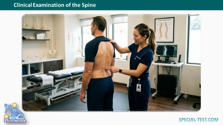
A meticulous clinical examination of the spine is essential for accurately localizing pathology and differentiating between mechanical, inflammatory, and neurogenic causes of spinal disorders.
1. History Taking
Pain Assessment
- Onset (Acute vs. Insidious):
- Acute onset is typical in post-traumatic injuries, sudden jerks, pyogenic infections (discitis), and tubercular infections.
- Insidious onset points toward mechanical low back pain, spondylitis, facet joint arthropathy, vascular structures, metastasis (unless precipitated by sudden trauma), primary bone tumors, and hemangiomas.
- Site:
- Axial (localized) pain at any spinal level often results from “mechanical” (non-neurogenic) structures like musculotendinous units, zygapophyseal (facet) joints, vertebrae, and the annulus fibrosus.
- Radiating pain indicates the involvement of neurological structures, commonly related to disc herniation, degenerative changes, neuroforaminal stenosis, space-occupying lesions, or intrinsic diseases of the cord/nerve roots (e.g., Herpes zoster).
- Nature of Pain:
- Throbbing, acute, unbearable pain suggests acute pyogenic infections or traumatic/pathological fractures (excluding mild osteoporotic fractures).
- Severe, constant pain may result from cauda equina syndrome, advanced tubercular infection, or acute cervical/dorsal/lumbar strains.
- Dull, constant aching pain is typically the result of chronic mechanical problems.
- Radiation Pattern: Both radiated (referred from another site) and radiating (traveling to another site) pain are common.The General Rule for Radiating Pain: It is the spinal level of the pathology—and not the exact anatomical structure involved—that determines the specific radiation pattern.
- Pain from the lumbar or cervical regions often radiates as a “lightning sensation” into the limbs, whereas dorsal segments radiate in a “band-like” fashion.
- Axially radiated pain can originate from visceral structures, including the aorta, carotid arteries, costovertebral/costotransverse articulations, pancreas, lungs, pleura, gallbladder, stomach, proximal duodenum, diaphragm, kidneys, ureters, and female pelvic organs.
- Non-spinal conditions presenting with radiating pain include lower trunk brachial plexopathies, peripheral neuropathies, cardiac conditions (heart/pericardium radiating to the upper limb), sacroiliac joint disorders, sacral stress fractures, and disorders of the ilia or hip joints.
- Diffuse vs. Localized: Localized pain often results from a focal injury to mechanical structures, whereas diffuse pain is typically radiated or referred.
- Aggravating and Relieving Factors:
- Spinal rotation and extension classically aggravate zygapophyseal (facet) joint pain.
- Forward flexion aggravates discogenic pain.
- Any spinal excursion (movement) is painful in inflammatory conditions of the spine.
- Neuroforaminal stenosis causes neurogenic claudication, which is triggered by exertion and relieved by forward flexion or sitting.
- Conversely, sitting and rising from a seated position worsen discogenic pain, while sleeping with the hips and knees flexed typically provides relief.
Trauma
- Document the exact energy dissipated from the impact.
- Note whether the mechanism was direct or indirect (e.g., heavy lifting, seat belt deceleration injuries).
Deformity
- Evaluate the onset, overall duration, and any sudden increase in size (which may indicate a vertebral collapse in pathological fractures).
- Note any association with pain, fever (suggestive of tuberculosis), or progressive stiffness (suggestive of ankylosing spondylitis).
Neurological Symptoms
- Assess for any loss of motor power, focal muscle weakness, or alterations in balance and gait.
Associated Medical & Systemic History
- Other Systems: Evaluate for genitourinary or gynecological complaints, signs of Marfan’s syndrome, ankylosing spondylitis, or vascular claudication.
- Past Medical History: Screen for Diabetes mellitus, tuberculosis, hypertension, hematological disorders, chronic pulmonary disorders, history of osteoporosis treatment, neurological disorders (e.g., epilepsy, Parkinsonism), and pre-operative HIV status.
- Personal History: Record smoking status, alcohol consumption, and any history of drug addiction.
2. Inspection
Attitude and Deformity
- Observe the patient’s natural posture (e.g., a rigid “military attitude”).
- Check the symmetry and levels of the shoulders.
- Perform a forward bending test to evaluate for scoliosis, kyphosis, or the spinal rigidity of ankylosing spondylitis.
- Perform a side bending test to look for a spinal list.
Lateral (Side) View Inspection
- Kyphotic Deformities: Normal thoracic kyphosis ranges between 21° and 33° (measured via Cobb’s method). Note the shape of any abnormal kyphosis:
- Knuckle: A sharp angular deformity involving a single vertebra.
- Gibbus: An angular deformity involving 2 to 3 vertebrae.
- Short angular/rounded kyphosis: Involves a small segment of the spine (4 to 8 vertebrae).
- Broad rounded kyphosis: Commonly seen in ankylosing spondylitis, senile osteoporosis, Scheuermann’s disease, and osteomalacia.
- Loss or partial reduction of kyphosis: Seen in early spinal tuberculosis due to paravertebral muscle spasm (“boarding”), flat back syndrome, or iatrogenic flattening following scoliosis surgical correction.
- Spinal Lordosis: (Average values: Lumbar lordosis = 40°–60°; Cervical lordosis = 20°–40°; Thoracic kyphosis = 20°–45°).
- Increased Lordosis: Commonly observed in females, obesity, spondylolisthesis, fixed flexion deformities of the hip, or as a constitutional variation or compensatory mechanism for increased thoracic kyphosis.
- Reduced Lordosis (Flattening/Reversal): Seen in the “sniffing position” of the cervical spine (face thrust anteriorly due to flexion at the cervico-thoracic junction and extension at upper segments, typical of ankylosing spondylitis), prolapsed intervertebral disc (PIVD), spinal infections, ankylosing spondylitis, and lumbar flatback syndrome (caused by osteoporotic/traumatic anterior wedging, advanced disc degeneration, or long thoracolumbar fusions).
Posterior (Back) View Inspection
- Alignment: Evaluate the position of the head relative to the pelvis. Drop a plumb line from the occiput (inion); in a straight spine, it must pass directly through the cleft of the buttocks.
- Anatomical Landmarks: Inspect the hairline, check for short neck or webbing, check shoulder levels, comparative positions of the scapular spines, and the inferior scapular angles.
- Deformities & Shifts:
- Look for a step-off deformity (indicative of spondylolisthesis).
- Identify any spinal list—an abrupt, planar shift of the spine to one side in the coronal plane without a definitive curvature, most commonly seen in the lumbar region.
- Pelvic Symmetry: Compare the heights of the iliac crests and the posterior superior iliac spines (dimples of Venus).
- Skin Markers: Carefully examine the skin for:
- Lumbar lipomas or localized hair patches (suggestive of spina bifida occulta).
- Port-wine stains (associated with spinal dysraphism/meningomyelocele).
- Café-au-lait spots and nodular skin swellings (Neurofibromatosis).
- Dermal hemangiomas, surgical scars, or active sinuses.
- Visible paravertebral muscle spasm or masses (e.g., meningomyelocele, paravertebral abscess, or lumbar hernia).
- Scoliosis Evaluation:
- Determine the sidedness of the deformity based on the direction of the lateral convexity (right vs. left).
- Adam’s Forward Bending Test: Have the patient bend forward to make structural deformities more prominent. Assess for structural rigidity, look for a “razor-back” rib hump, and identify the apex of the primary curve by locating the zone of maximum vertebral rotation.
- Lateral Bending: Evaluate the flexibility and correctability of the spinal curves. Note that vertebral rotation is structurally fixed in primary curves; other curves are typically flexible, compensatory curves.
Anterior (Front) View Inspection
- Ensure the head is seated squarely over the shoulders with the chin aligned directly over the sternal notch.
- Inspect the alignment of the hyoid bone, thyroid cartilage, and sternocleidomastoid muscles.
- Examine the sternum for chest wall deformities like pectus excavatum (funnel chest) or pectus carinatum (pigeon chest).
- Check the position and symmetry of the umbilicus.
Gait Analysis
- Observe the patient’s walking pattern to identify specific neurological or antalgic gaits:
- Shuffling gait (posterior cord syndrome)
- Slapping/high-stepping gait (foot drop/peroneal nerve injury)
- Broad-based/halting gait (sensory or cerebellar ataxia)
- Festinating gait (Parkinsonian features)
- Antalgic gait (pain-avoiding gait cycle)
- Functional Gait Tests:
- Heel walking: Specifically tests the ankle dorsiflexors (primarily the L5 nerve root).
- Toe walking: Specifically tests the ankle plantarflexors (primarily the S1 nerve root).
3. Palpation and Percussion
Palpation Protocol
- Local Temperature: Assess for any localized warmth that may indicate active inflammation or infection.
- Tenderness Localization:
- Superficial tenderness: Elicited with light direct pressure, often indicating cutaneous or subcutaneous affections.
- Deep tenderness: Elicited by applying firm, direct pressure directly over the spinous processes.
- Twist (Rotator) Tenderness: Apply lateral pressure to the side of the spinous process in an attempt to induce micro-rotation of the vertebra.
- Thrust Tenderness: Elicited by gently thrusting or percussing the spine using a closed fist.
- Spinous Process Palpation: Palpate sequentially from the inion down to the coccyx. Pay special attention to palpable landmarks (C2, C7, T1, T12).
- Identify structural defects (spina bifida), bony thickening, or iatrogenic defects (prior laminectomy margins).
- Assess alignment and abnormal prominence. Note: The upper spinous process becomes abnormally prominent in conditions causing anterior wedging or distraction of the posterior elements (e.g., traumatic, osteoporotic, or tubercular fractures, and retrolisthesis). Conversely, the lower spinous process appears prominent when there is a forward subluxation of the superior spine, as seen in fracture-dislocations and spondylolisthesis.
- Soft Tissues & Anterior Palpation: Palpate the paraspinal gutters and interspinous regions for supraspinous ligament gaps or fluctuant masses (such as a cold Pott’s abscess). From the front, palpate the carotid tubercle (Chassaignac’s tubercle at C6), the sternal notch, and any anterior chest wall deformities.
Percussion
- Perform direct percussion over the spinous processes to screen for deep osseous pain (e.g., infection or tumor).
- Percuss the costotransverse joints to evaluate for localized thoracic segment pathology.
4. Range of Motion (Moving)
- Flexion:
- Cervical Spine: Instruct the patient to touch their chin directly to their chest.
- Lumbar Spine: Instruct the patient to bend forward with straight knees. Measure either the angle of the trunk relative to the vertical plane or the finger-to-floor distance (Normal values: 90° flexion or less than 10 cm finger-to-floor distance).
- Extension:
- Cervical Spine: Instruct the patient to look directly up at the ceiling.
- Lumbar Spine: Instruct backward bending (Normal range: 20°–30°). This movement is markedly limited and painful in patients presenting with facet arthropathy or neuroforaminal narrowing, as extension further decreases the neuroforaminal cross-sectional area.
- Lateral Rotation:
- Evaluate right and left rotation in the cervical spine (Normal range: ~80°).
- To evaluate lumbar spine rotation independently, have the patient sit on a stool to lock the pelvis, and measure the angle achieved between the shoulder girdle plane and the pelvic plane.
- Lateral Bending:
- Evaluate thoracic/lumbar lateral bending by tracking the floor-to-fingertip distance as the patient slides their hand down the lateral thigh.
- Thoracic Spine Isolation: Because true thoracic flexion and extension are minimal due to rib cage splinting, evaluate these motions by having the patient sit on a straight, flat-backed wooden chair while attempting to bend forward and backward. Detailed structural tracking can be supplemented via direct segment skin measurement.
5. Neurological Examination
A structured neurological assessment is vital to correlate the structural findings from your inspection and palpation with functional nerve root or spinal cord status.
Motor Evaluation by Nerve Root
Testing specific muscle groups helps localize the exact level of nerve root compression or pathology:
- C5: Shoulder abduction (Deltoid) and elbow flexion (Biceps brachii).
- C6: Wrist extension (Extensor carpi radialis longus/brevis).
- C7: Elbow extension (Triceps brachii) and wrist flexion.
- C8: Finger flexion (Flexor digitorum profundus to the middle finger).
- T1: Finger abduction and adduction (Interossei).
- L2: Hip flexion (Iliopsoas).
- L3: Knee extension (Quadriceps femoris).
- L4: Ankle dorsiflexion (Tibialis anterior).
- L5: Great toe extension (Extensor hallucis longus).
- S1: Ankle plantarflexion (Gastrocnemius and soleus).
Reflex Assessment
Document deep tendon reflexes (DTRs) to differentiate between Upper Motor Neuron (UMN) lesions (hyperreflexia, clonus) and Lower Motor Neuron (LMN) lesions (hyporeflexia or arreflexia):
- Biceps Reflex: Evaluates the C5 nerve root.
- Brachioradialis Reflex: Evaluates the C6 nerve root.
- Triceps Reflex: Evaluates the C7 nerve root.
- Knee Jerk (Patellar Reflex): Evaluates the L4 nerve root (with some L3 contribution).
- Ankle Jerk (Achilles Reflex): Evaluates the S1 nerve root.
- Pathological Reflexes: Always screen for the Babinski sign and Hoffmann’s sign to rule out cervical or thoracic myelopathy.
6. Special Clinical Tests
These provocative maneuvers are designed to stress specific anatomical structures to confirm your clinical suspicions.
Cervical Spine Tests
- Spurling’s Test (Foraminal Compression Test): With the patient seated, extend and rotate the head toward the affected side, then apply a gentle downward axial force on the top of the head.
- Positive result: Radiation of pain into the ipsilateral arm, indicating cervical radiculopathy.
- Distraction Test: Place one hand under the patient’s chin and the other under the occiput, then gently lift the head vertically.
- Positive result: Relief or significant reduction of radicular symptoms, confirming cervical nerve root compression.
- Lhermitte’s Sign: Have the patient actively or passively flex the cervical spine forward.
- Positive result: An electric shock-like sensation traveling down the spine into the limbs, indicating cervical myelopathy, multiple sclerosis, or severe cord compression.
Lumbar Spine & Radicular Stress Tests
- Straight Leg Raising Test (SLRT) / Lasègue’s Sign: With the patient supine, passively lift the extended leg on the affected side by the heel. Note the angle at which pain occurs (typically positive between 30° and 70°).
- True Positive: Sharp, shooting radicular pain extending below the knee along the distribution of the sciatic nerve (L4, L5, S1 roots). Localized back or hamstring pain does not constitute a positive test.
- Bragard’s Sign: Lower the leg slightly from the point of pain and acutely dorsiflex the foot. Re-precipitation of radicular pain confirms dural tension.
- Crossed (Contralateral) SLRT: Perform the SLRT on the unaffected leg.
- Positive result: Radicular pain is reproduced in the opposite (symptomatic) leg. This is highly specific for a large, central, or extruded disc herniation.
- Femoral Nerve Stretch Test (Reverse SLRT): Place the patient in a prone position. Flex the knee to 90° and passively extend the hip.
- Positive result: Pain radiating down the anterior aspect of the thigh, indicating irritation of the upper lumbar nerve roots (L2, L3, L4).
Sacroiliac Joint (SIJ) & Hip Differentiating Tests
- FABER (Patrick’s) Test: Flex, Abduct, and Externally Rotate the hip so the lateral malleolus rests on the opposite knee. Apply downward pressure on the flexed knee while stabilizing the opposite iliac crest.
- Interpretation: Anterior groin pain indicates a hip joint issue; posterior pain localized to the SI region indicates sacroiliac joint pathology.
- Gaenslen’s Test: Have the patient lie supine at the edge of the examination table. Instruct them to hug one knee tightly to their chest while the opposite leg hangs completely off the edge of the table into hyperextension.
- Positive result: Pain localized to the sacroiliac joint on the extended leg side, indicating SIJ inflammation or mechanical dysfunction.
7. Conclusion & Documentation
Once the physical examination is complete, synthesize your findings into a clear clinical picture. Correlate the systemic history (e.g., insidious onset, night pain, weight loss) with your physical findings (e.g., rigid spine, localized deep tenderness, or focal neurological deficits). This clinical framework forms the foundation for targeted diagnostic imaging, such as standard radiographs, Cobb angle measurements for spinal deformities, or advanced magnetic resonance imaging (MRI).