Knee Examination: History Taking and Clinical Examination
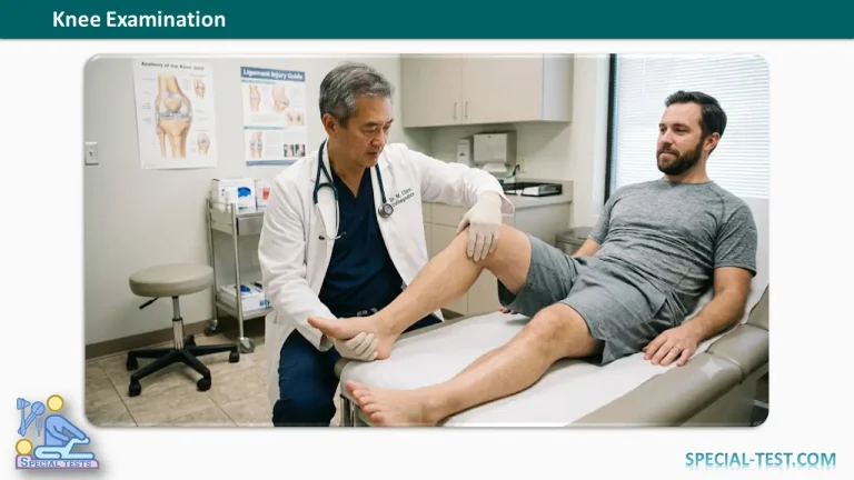
The clinical knee examination of the knee is one of the most important components of orthopedic assessment. A structured approach helps identify the underlying pathology, distinguish between traumatic and non-traumatic conditions, and determine whether the problem originates from the knee or is referred from another region such as the hip or lumbar spine.
A complete knee assessment consists of:
- History taking
- Inspection
- Palpation
- Assessment of range of motion
- Special orthopedic tests
- Neurovascular examination (when indicated)
History Taking
A detailed history often provides the diagnosis before physical examination begins.
Pain
Assess the characteristics of the patient’s pain carefully.
- Onset
- Sudden (trauma, ligament injury, infection)
- Gradual (osteoarthritis, inflammatory arthritis)
- Duration
- Acute
- Chronic
- Recurrent
- Severity
- Mild
- Moderate
- Severe
- Character
- Aching pain → degenerative disorders, bone tumors
- Throbbing pain → infection
- Progression
- Insidious onset → degenerative or mechanical disorders
- Acute onset → trauma or septic arthritis
- Diurnal variation
- Morning stiffness → inflammatory arthritis
- Evening pain → mechanical disorders or osteoarthritis
- Night pain → inflammatory disease, infection, tuberculosis, or malignancy
- Activity-related pain
- Pain that worsens with activity commonly suggests osteoarthritis or mechanical pathology.
- Radiation
- Pain radiating to the calf may occur with degenerative or mechanical disorders.
- Consider referred pain from the lumbar spine.
- Pain after prolonged sitting (“Theater Sign”)
- Suggests patellofemoral disorders.
- Bar or vice-like anterior knee pain
- May indicate patella baja (low-riding patella).
- Pain elsewhere
- Hip pathology
- Lumbar spine disorders
- Inflammatory polyarthritis
- Previous episodes
- Previous injuries
- Prior surgery
- Recurrent instability
Deformity
Ask whether the patient has noticed any deformity.
Common deformities include:
- Genu varum
- Osteoarthritis
- Rickets
- Post-traumatic deformity
- Genu valgum
- Rheumatoid arthritis
- Rickets
- Genu recurvatum
- Poliomyelitis
- Generalized ligamentous laxity
- Flexion deformity
- Infection
- Joint effusion
- Hemophilia
- Scarring
- Triple deformity
- Tuberculosis
- Broadening of the knee
- Osteophytes
- Hemophilic arthropathy
Patellar abnormalities include:
- Patella alta
- Patella baja
- Lateral patellar subluxation
- Bipartite patella
- Small or hypoplastic patella
Also enquire about deformities involving:
- Hip
- Foot
- Small joints of the hands
These may indicate systemic disease.
Swelling
Determine:
- Onset
- Duration
- Mechanism
- Traumatic
- Atraumatic
- Associated symptoms
- Fever
- Pain
- Weight loss
Ask whether aspiration has been performed previously.
The appearance of aspirated fluid can provide important diagnostic clues.
| Aspirate | Possible diagnosis |
|---|---|
| Straw-colored | Osteoarthritis, inflammatory arthritis, synovitis |
| Purulent | Septic arthritis |
| Blood-stained | Hemarthrosis, trauma, hemophilia |
| Brown or amber | Pigmented villonodular synovitis (PVNS) |
Long-standing painless swellings may represent:
- Meniscal cyst
- Osteochondroma
- Benign soft tissue tumors
Painful swellings raise concern for:
- Infection
- Malignancy
- Hematoma
Instability (Laxity)
Ask specifically whether the knee:
- “Gives way”
- “Goes out”
- Buckles unexpectedly
These symptoms may suggest:
- ACL rupture
- Patellar instability
- Meniscal pathology
- Loose bodies
- Articular cartilage injury
- Quadriceps weakness
- Neuromuscular disease
- Generalized ligamentous laxity
Locking
Determine whether locking is:
True Locking
Characterized by inability to fully extend the knee.
Common causes include:
- Meniscal tear
- Loose bodies
- Torn ACL stump
False Locking (Catching)
Usually caused by:
- Patellofemoral disorders
- Hoffa’s fat pad impingement
Limitation of Movement or Stiffness
Determine whether restriction is:
Painful
Suggests:
- Arthritis
- Trauma
- Infection
- Tumor
Painless
Usually mechanical, including:
- Muscle contracture
- Tendon contracture
- Fascial tightness
- Malunited fracture
- Osteophytes
- Arthrodesis
Restriction may also be:
- Intra-articular
- Extra-articular
Other Important Symptoms
Ask about:
- Crepitus
- Clicking
- Clunking
- Snapping
- Catching sensations
- Previous surgery
- Previous injections
- Functional limitations
- Sports participation
Patient-reported outcome measures such as the International Knee Documentation Committee (IKDC) Score may also be useful during evaluation.
Inspection
Inspection should begin with the patient standing.
Standing Examination
Kneel in front of the patient to inspect the knees at eye level.
Attitude
Observe:
- Knee extension
- Hip extension
- Plantigrade feet
- Patellae facing forward
Alignment
Evaluate overall lower limb alignment.
Look for:
- Normal physiological alignment
- Genu valgum
- Genu varum
- Windswept deformity
Inspect the Knee from All Directions
Look for:
Swelling
Generalized or localized swelling may indicate:
- Bursitis
- Meniscal cyst
- Baker’s cyst
- Osteochondroma
- Osgood-Schlatter disease
- Sinding-Larsen-Johansson syndrome
- Patellar tendinopathy (Jumper’s knee)
- Hoffa’s disease
Joint Effusion
Observe for fullness around the patella.
Skin Changes
Inspect for:
- Erythema
- Sinuses
- Surgical scars
- Neurofibromatosis
- Psoriatic lesions
Patellar Abnormalities
Assess for:
- Bipartite patella
- Squinting patella
- Frog-eye patella
- Patella magna
- Patella breva
Muscle Wasting
Particularly examine:
- Vastus medialis obliquus (VMO)
Quadriceps wasting commonly accompanies chronic knee pathology.
Gait Examination
Observe walking from:
- Front
- Back
- Side
Frontal Plane
Look for:
- Valgus thrust
- Varus thrust
- Circumduction gait
- Varus recurvatum thrust
- Duck-footed gait
Lateral View
Observe for:
- Antalgic gait
- Stiff-knee gait
- Flexed-knee gait
Sitting Examination
Assess:
- Patella alta
- Patella baja
- J-sign
- Dynamic patellar tracking
- Patellar tilt
Supine Examination
Reassess findings noted during standing.
Pay particular attention to:
- Flexion deformity
- Joint effusion
- Alignment correction
Correction of deformity in the supine position often indicates an intra-articular cause.
Palpation
Always compare both knees.
Anterior Aspect
Temperature
A warm knee suggests:
- Infection
- Inflammatory arthritis
- Active synovitis
Joint Effusion
Assess using:
- Fluid shift test (≈15 mL)
- Cross-fluctuation test (≈30 mL)
- Patellar tap (ballottement)
- Transillumination (selected cases)
Palpate the synovium for thickening or bogginess.
Patella and Extensor Mechanism
Examine:
- Patellar facet tenderness
- Patellar tendon
- Quadriceps tendon
- Bipartite patella
- Tibial tubercle
Assess:
- Patellar glide
- Patellar tracking
- Patellar tilt
- Patellar grind test
- Fairbank apprehension test
Wilson Test
Wilson’s test helps detect osteochondritis dissecans of the medial femoral condyle.
Medial Aspect
Palpate:
- Medial joint line
- Pes anserinus
- Pes anserine bursa
- Meniscal cyst
- Medial collateral ligament (MCL)
Joint line tenderness is highly suggestive of a medial meniscal tear.
Lateral Aspect
Assess:
- Lateral joint line
- Lateral collateral ligament (LCL)
- Iliotibial band
- Gerdy’s tubercle
Perform:
- Ober’s test (iliotibial band tightness)
- Allis (Galeazzi) test when limb length discrepancy is suspected
Posterior Aspect
Examine the patient prone when appropriate.
Assess:
- Flexion deformity
- Baker’s cyst
- Popliteal masses
- Popliteal aneurysm
- Enlarged lymph nodes
Craig’s (Ryder) test may be used to assess femoral anteversion.
Clinical Pearls
- The patient’s history frequently provides the diagnosis before examination.
- Always compare the affected knee with the contralateral side.
- Observe gait before palpation to avoid altering the patient’s natural walking pattern.
- Inspect the patient both standing and supine, as subtle deformities or effusions may only become apparent in one position.
- A systematic examination reduces the likelihood of missing significant pathology.
References
- Hoppenfeld S. Physical Examination of the Spine and Extremities.
- Magee DJ. Orthopedic Physical Assessment.
- Solomon L, Warwick D, Nayagam S. Apley’s System of Orthopaedics and Fractures.
- American Academy of Orthopaedic Surgeons (AAOS). Clinical evaluation of the knee.
- Norris R, et al. The clinical examination of the knee. BMJ. 2018.
- Logerstedt DS, et al. Knee stability and movement coordination impairments. Journal of Orthopaedic & Sports Physical Therapy (JOSPT). 2017.
- American College of Rheumatology recommendations for the evaluation of knee pain and arthritis.