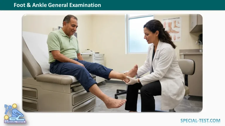
Foot & Ankle General Examination
History Taking
Age
- Congenital talipes equinovarus (CTEV) is present since birth.
- Talipes equinovarus (TEV) secondary to polio, neural tube defects, and other neurological conditions develops later.
- Congenital vertical talus (CVT) is usually recognized when the child begins walking (around 1 year of age).
Sex
- CTEV is more common in boys.
Pain
Assess the following characteristics:
- Duration
- Site
- Radiation
- Type
- Character
- Aggravating factors
- Relieving factors
- Diurnal variation
- Postural variation
Swelling
Evaluate:
- Duration
- Onset (following trauma, fever, involvement of other joints, morning stiffness)
- Progression
- Progressive enlargement (suggests tumors)
- Regressive swelling (common after trauma)
- Intermittent swelling (may indicate infection)
- Aggravating factors (e.g., walking in subtalar arthritis)
- Relieving factors (e.g., antibiotics for infection, chemotherapy for tumors)
- Response to previous treatment
- Diurnal and postural variation
- Presence of deformity in the opposite foot
Limp
Determine:
- Onset
- Duration
- Painful or painless
- Progressive or non-progressive
Instability
Assess:
- Duration
- Onset (especially after trauma)
- Unilateral or bilateral involvement (may indicate generalized ligamentous laxity)
- Whether instability occurs on even or uneven surfaces (common in a stiff subtalar joint)
Deformity
Evaluate:
- Time of onset
- Present at birth (CTEV)
- Appeared later (acquired clubfoot)
- Around walking age (CVT)
- Following febrile illness with muscle weakness (polio)
- Progression
- Congenital deformities are generally less progressive than acquired deformities.
- Previous treatment
- Casting
- Surgery
- Response to treatment
Associated Diseases
- Previous history of fever, myalgia, and limb weakness suggestive of poliomyelitis.
Inspection
Gait
Observe for:
- Antalgic gait
- Short-limb gait
- Foot drop
- Equinus gait
- Stiff first metatarsophalangeal (MTP) joint
Anterior Aspect
- Alignment
- Hallux valgus or hallux varus
- Claw, hammer, or mallet toes
- Relationship of the forefoot, midfoot, and hindfoot
- Tibia vara
- Tibial rotation
- Skin
- Discoloration
- Ulcers
- Dilated veins
- Edema (pitting or non-pitting)
- Toes
- Loss of transverse skin creases (may occur in polio)
- Corns (heloma durum)
- Nail deformities from fungal infection
- Paronychia
- Ingrown toenail
- Bony Prominences
- Medial osteophyte over the first MTP joint (bunion)
- Lateral osteophyte over the fifth MTP joint (bunionette)
- Extensor Tendons
- Observe extensor hallucis longus (EHL) and extensor digitorum longus (EDL) during active contraction.
Mnemonic: The Himalayas Are Not Dry Places
- Tibialis Anterior
- Extensor Hallucis Longus
- Anterior Tibial Artery
- Deep Peroneal Nerve
- Extensor Digitorum Longus
- Peroneus Tertius
- Malleoli Relationship
- Normally the lateral malleolus lies lower and more posterior than the medial malleolus.
- Malleolar Swelling
- Trauma
- Tendinitis
- Anterior Tibia
- Swelling
- Deformity
Lateral Aspect
Inspect:
- Lateral malleolus
- Base of the fifth metatarsal
- Achilles tendon
- Peroneus brevis tendon
- Any swelling or deformity
Posterior Aspect
- Hindfoot varus or valgus
- Too Many Toes Sign
- More than two toes visible from behind indicates forefoot abduction, commonly associated with pes planus.
- Heel
- Width
- Shape
- Position
- Ask the patient to stand on tiptoes.
- Assess the windlass mechanism (arch elevation and hindfoot inversion).
- Examine:
- Plantar fat pad
- Calcaneal tuberosity
- Haglund deformity (pump bump)
- Retrocalcaneal bursa
- Look for bursitis.
- Achilles tendon
- Tendinitis
- Rupture (typically 2–6 cm proximal to insertion)
- Swelling
- Calf Muscle
- Compare for muscle wasting, which may occur in:
- Residual CTEV
- Achilles tendon rupture
- Prolonged immobilization
Medial Aspect
- Medial longitudinal arch
- Pes cavus
- Pes planus
- Rocker-bottom deformity
- Bony landmarks
- Medial malleolus
- First metatarsal head
- Calcaneal tuberosity
- Navicular tuberosity
- Tibialis posterior tendon
Mnemonic: The Doctors Are Never Happy
- Tibialis Posterior
- Flexor Digitorum Longus
- Posterior Tibial Artery
- Posterior Tibial Nerve
- Flexor Hallucis Longus
Plantar Aspect
- Callosities indicating abnormal weight-bearing
- Painful metatarsal calluses
- Corns (hard or soft)
- Ulcers (especially diabetic ulcers)
- Plantar warts
- Tinea pedis
Palpation
Anterior
- Local temperature
- Tenderness over:
- Tibial crest (stress fracture)
- Talar dome (osteochondral lesion)
- Navicular (Köhler disease)
- Talonavicular joint
- Cuneiforms
- Metatarsals
- First MTP joint
- Second MTP joint (Freiberg disease)
- Morton neuroma tenderness between the third and fourth metatarsal heads
- Swelling
- Joint effusion
- Extensor tendons
- Corns and ingrown toenails
- Tinel sign over the deep peroneal nerve (anterior tarsal tunnel syndrome)
Lateral
Palpate:
- Lateral malleolus
- Anterior talofibular ligament
- Calcaneofibular ligament
- Peroneal tendons
- Calcaneus
- Calcaneocuboid joint
- Sinus tarsi
- Fibular shaft
Posterior
- Gastrocnemius-soleus complex
- Achilles tendon
- Retrocalcaneal bursa
Medial
Palpate:
- Medial malleolus
- Subcutaneous tibial border
- Talar head
- Navicular tuberosity
- Flexor hallucis longus
- Flexor digitorum longus
- Tibialis posterior tendon
- Tinel sign over the posterior tibial nerve and plantar nerves
Plantar
Assess:
- Tender callosities
- Sesamoid tenderness
- Plantar fascia tenderness
- Plantar fibromatosis nodules
- Plantar fat pad tenderness
Range of Motion
Ankle
Evaluate:
- Dorsiflexion: 20°
- Plantar flexion: 50°
Assess passive dorsiflexion with the knee both extended and flexed when equinus deformity is present.
Interpretation:
- Increased dorsiflexion with knee flexion → isolated gastrocnemius tightness.
- No change → isolated soleus contracture.
- Partial improvement but persistent limitation → combined gastrocnemius and soleus tightness.
Subtalar Joint
Assess:
- Inversion: 40°
- Eversion: 20°
The examination is best performed with the patient prone while stabilizing the talus.
Forefoot
Evaluate:
- Abduction
- Adduction
The calcaneus should be maintained in a neutral position.
Great Toe
Metatarsophalangeal (MTP) Joint:
- Extension: 70°
- Flexion: 45°
Interphalangeal (IP) Joint:
- Flexion: 90°
- Extension: 0°
Lesser Toes
Assess:
- Flexion and extension at the interphalangeal joints (40°)
- Flexion (40°) and extension (0°) at the MTP joints
- Toe abduction
- Toe adduction toward the second toe