Elbow General Examination
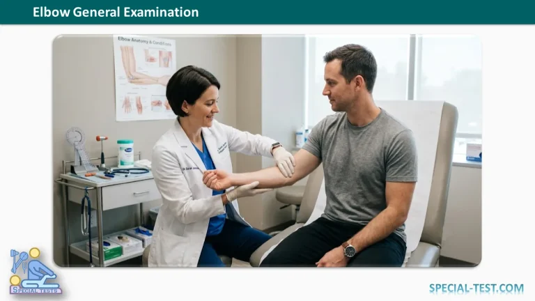
History Taking
A detailed history is the first step in evaluating a patient with elbow pain or dysfunction. It helps differentiate traumatic, inflammatory, degenerative, neurological, and infectious conditions.
Pain
Assess the characteristics of the pain by asking about:
- Onset: Sudden or gradual.
- Location:
- Bony pain: Medial epicondyle, lateral epicondyle, supracondylar region, olecranon, radial head, or joint line.
- Soft tissue pain: Common extensor origin, common flexor origin, bursae, tendons, or muscle belly.
- Duration: Acute or chronic.
- Radiation: Pain extending to the forearm, wrist, shoulder, or hand.
- Severity and character: Sharp, dull, aching, burning, or throbbing.
- Associated symptoms:
- Fever (suggesting septic arthritis or osteomyelitis)
- Trauma
- Locking or clicking
- Instability
- Morning stiffness
- Numbness or tingling (possible ulnar nerve involvement)
- Aggravating and relieving factors: Activities, lifting, gripping, sports, or rest.
Swelling
Evaluate the swelling by determining:
- Time of onset and duration.
- Localized or diffuse swelling.
- Site of swelling:
- Olecranon bursa
- Lateral soft spot (Anconeus triangle)
- Medial aspect
- Generalized around the joint
- Associated redness, warmth, pain, fever, or previous injury.
Limitation of Movement
Determine whether movement restriction is associated with:
- Trauma
- Joint swelling
- Pain
- Previous immobilization
- Manipulation or massage
- Infection
- Previous surgery or other iatrogenic causes
Deformity
Ask whether the deformity is:
- Congenital or acquired.
- Related to previous trauma.
- Associated with infection or inflammatory disease.
- Secondary to treatment or surgery.
- Due to neuropathic (Charcot) arthropathy.
Additional History
Important additional questions include:
- Previous fractures or elbow dislocations.
- Recurrent instability.
- History of haemophilia or bleeding disorders.
- Previous corticosteroid injections.
- Occupation and sporting activities (especially throwing sports or racquet sports).
- Previous attempts at manipulation or traditional massage.
- Systemic diseases such as rheumatoid arthritis, gout, diabetes, or tuberculosis.
Inspection
Inspection should be performed with both upper limbs fully exposed and compared simultaneously.
General Observation
Observe for:
- Patient posture.
- Attitude of the elbow.
- Muscle wasting.
- Skin changes.
- Scars.
- Sinuses.
- Bruising.
- Erythema.
- Obvious deformity.
Attitude
Assess:
- Carrying angle of both elbows.
- Flexion deformity.
- Fixed extension or flexion posture.
- Varus or valgus deformity.
Anterior (Front) View
Inspect:
- Flexion crease (normally 1–2 cm proximal to the joint line at the level of the interepicondylar axis).
- Medial and lateral epicondyles.
- Biceps tendon.
- Lacertus fibrosus.
- Common flexor origin.
- Common extensor origin.
- Swelling.
- Enlarged epitrochlear lymph nodes.
Lateral View
Look for:
- Fullness over the Anconeus triangle (soft spot).
- Swelling suggestive of elbow joint effusion.
- Common extensor origin.
- Olecranon process.
- Triceps tendon prominence.
- Olecranon bursa.
- Radial head prominence or dislocation.
- Biceps muscle contour.
Medial View
Inspect:
- Medial epicondyle.
- Supracondylar depressions.
- Common flexor origin.
- Soft tissue swelling.
Posterior View (Elbow Flexed to 90°)
Observe:
- Olecranon process.
- Triceps muscle bulk.
- Paraolecranon fossae.
- The normal three-point relationship formed by:
- Medial epicondyle
- Lateral epicondyle
- Olecranon
Disruption of this relationship may indicate elbow dislocation or distal humeral fracture.
Palpation
Palpation should begin away from the painful area and proceed systematically.
Superficial Palpation
Assess:
- Local temperature.
- Generalized tenderness.
- Skin texture.
- Fluctuation or edema.
Deep Palpation
Bony Landmarks
Palpate:
- Medial epicondyle.
- Lateral epicondyle.
- Olecranon.
- Olecranon fossae.
- Radial head.
- Capitellum (important in osteochondritis or Panner disease).
- Supracondylar ridges.
Soft Tissue Structures
Palpate:
- Common extensor tendon.
- Common flexor tendon.
- Triceps tendon.
- Distal biceps tendon.
- Olecranon bursa.
- Anconeus triangle.
- Any palpable muscle masses (e.g., myositis ossificans).
Assessment of Swellings
For any swelling, evaluate:
- Site.
- Size.
- Shape.
- Surface.
- Margins.
- Consistency.
- Tenderness.
- Mobility.
- Compressibility.
- Reducibility.
- Pulsatility.
- Fluctuation.
- Transillumination (if appropriate).
Supracondylar Ridges
Palpate both medial and lateral supracondylar ridges simultaneously to identify:
- Irregularity.
- Thickening.
- Cortical discontinuity.
- Supracondylar spur.
- Loss of normal contour.
Three-Point Relationship
Confirm preservation of the normal relationship between:
- Olecranon.
- Medial epicondyle.
- Lateral epicondyle.
Ulnar Nerve
Palpate behind the medial epicondyle for:
- Thickening.
- Tenderness.
- Subluxation during elbow flexion.
- Tinel’s sign.
Joint Line
Palpate circumferentially for:
- Tenderness.
- Synovial thickening.
- Joint effusion.
Vascular Examination
Assess:
- Brachial artery pulsation.
- Radial pulse.
- Ulnar pulse.
- Capillary refill.
- Distal limb temperature.
Range of Motion
Both active and passive movements should be examined and compared with the opposite side.
Flexion
- Normal: 0° to 140°
Extension
- Returns the elbow to the neutral position.
- Physiological hyperextension up to 10° may be present, particularly in females and children.
Forearm Supination
- Normal: Approximately 85°
Forearm Pronation
- Normal: 70–80°
During Movement, Assess
- Pain.
- Crepitus.
- Mechanical locking.
- Instability.
- Muscle spasm.
- Terminal restriction.
- End feel.
Special Clinical Considerations
The elbow examination should always include assessment of adjacent joints:
- Shoulder
- Wrist
- Cervical spine
A focused neurovascular examination is also essential, including:
- Motor function
- Radial nerve
- Median nerve
- Ulnar nerve
- Sensory examination
- Peripheral vascular status
Failure to examine these structures may result in missed diagnoses, particularly in traumatic injuries.
Clinical Pearls
- Loss of the normal three-point relationship usually indicates elbow dislocation or displaced supracondylar fractures.
- Tenderness over the lateral epicondyle suggests lateral epicondylitis (tennis elbow).
- Tenderness over the medial epicondyle suggests medial epicondylitis (golfer’s elbow).
- Swelling over the olecranon is characteristic of olecranon bursitis.
- Pain over the radial head with limited forearm rotation may indicate a radial head fracture.
- Ulnar nerve subluxation or tenderness behind the medial epicondyle may indicate cubital tunnel syndrome.
References
- Hoppenfeld S. Physical Examination of the Spine and Extremities. Appleton & Lange.
- Magee DJ. Orthopedic Physical Assessment. Elsevier.
- Campbell’s Operative Orthopaedics. Elsevier.
- American Academy of Orthopaedic Surgeons (AAOS). Examination of the Elbow.
- National Center for Biotechnology Information (NCBI Bookshelf). Clinical Methods: The History, Physical, and Laboratory Examinations.