Derbolowsky Test
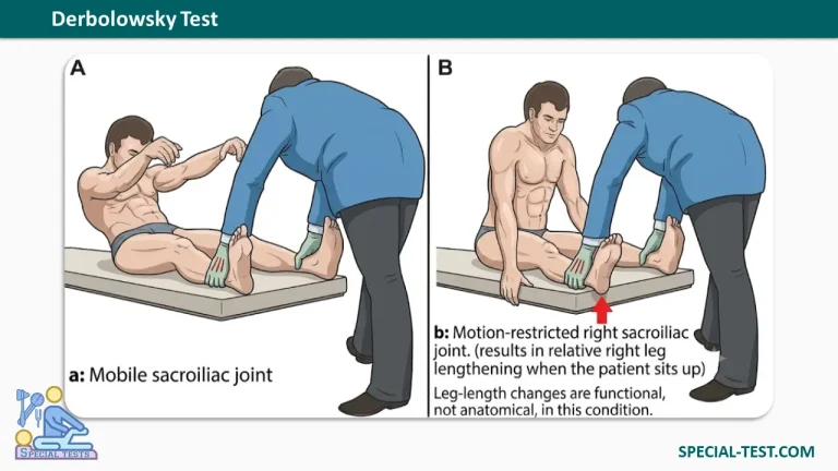
What is the "Derbolowsky Test" used for?
- Derbolowsky test is a clinical test used to assess a leg length difference: an advancement phenomenon with the patient supine.
How to perform the "Derbolowsky Test"?
- The patient is supine.
- The examiner grasps both ankles, palpates the patient’s medial malleoli with each thumb, and evaluates the relative level and rotation of the medial malleoli using the positions of the thumbs as reference.
- The patient is asked to sit up; either the examiner may help the patient do so, or the patient may use his or her hands for support.
- The legs should be lifted off the table to prevent interference.
- Then the level and rotation position of the malleoli are again evaluated.
- Lastly, the patient is asked to bend the trunk maximally forward to come as close to the extended knees as possible.
- The derbolowsky test should be carried out several times to prevent false-positive test results due to muscle tension.
What is the positive result?
- Forward advancement in the supine position suggests pelvic rotation.
- Where there is a motion restriction in the sacroiliac joint without any play between the sacrum and ilium, the ipsilateral leg will be longer when the patient sits up and apparently shorter or the same length as the other leg when the patient is supine.
- The examiner measures the difference in the level of the two malleoli, which previously were at the same level.
The differential diagnosis should consider whether something other than a motion restriction in the sacroiliac joint may be causing the variable leg length difference.
- Possible such causes include shortening of the hamstrings or genuine anatomical leg lengthening or shortening.
- Pain during the test could suggest loosening of the sacral structure, muscular foreshortening, or neurologic pain from a protruding or herniated disk.
- Derbolowsky test is considered to be diagnostically significant if the difference in levels amounts to at least 1 to 2 cm.
- When one sees larger differences in connection with myalgic pain, then one should consider shortened hamstring muscles as a cause.
- If the difference is greater than 5 cm combined with symptoms of radicular pain and pelvic rotation with compensatory flexion of the knee, then one should consider vertebral disk dysfunction.
References
- Clinical Tests for the Musculoskeletal 3rd Ed. Book