Beevor Sign
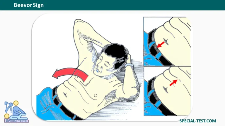
What is the "Beevor Sign" used for?
- Beevor Sign is used to evaluate the rectus abdominis muscle weakness or paralysis due to spinal cord lesions between T10-12.
How to perform the "Beevor Sign"?
- The patient is positioned supine, with the knees flexed and both feet flat on the bed.
- The patient is asked to raise the head against resistance, cough, or attempt to sit up with the hands resting behind the head.
- The clinician observes for motion at the umbilicus, which should remain in a straight line.
What is the positive result?
- The umbilicus shifts to the side of stronger musculature. Superior shift implicates weakness of muscles innervated by lower thoracic spine whereas inferior shift implicates upper thoracic spine weakness at or below T7.
- Positive Beevor sign, an upward deflection of the umbilicus on flexion of the neck, is the result of paralysis of the inferior portion of the rectus abdominis muscle, so that the upper fibers predominate, pulling the umbilicus upwards.
- False positive test occurs when the abdominal musculature is extensively weak, the thorax may pull away from the pelvis and arching of back occurs causing tautness of the anterior abdominal muscles mimicking actual contraction of the muscles.
Beevor sign is a common finding in patients with facioscapulohumeral dystrophy, even before functional weakness of abdominal wall muscles is apparent, but is absent in patients with other facioscapulohumeral disorders.
Beevor sign can be present in the following conditions:
- Spinal cord lesion between T10 and T12 segment, tumors, for instance. Noteworthy, reports exist of acute Beevor sign with spinal cord infarction due to vascular lesion below T10.
- Facioscapulohumeral muscular dystrophy (FSHD) is autosomal dominant muscle dystrophy. Beevor sign is considered as a “sine qua non” clinical sign of this disease. Although some authors reported that this sign is 90% sensitive and specific for FSHD, other researchers believe it is specific (over 90%) but less sensitive (54%) and can help in diagnosis. Furthermore, it is found more frequently in typical than atypical FSHD.
There are less frequent reports for this sign in the following diseases:
- Pompe disease: type 2 glycogen storage disease.
- GNE myopathy (autosomal recessive myopathy): this sign was observed in 90% of the patients in one study.
- Tubular aggregate myopathy.
- Myotonic dystrophy.
- Sporadic inclusion body myositis (IBM).
- Amyotrophic lateral sclerosis.
- Acid maltase deficiency in an adult patient.
References
- Post M: Physical Examination of the Musculoskeletal System. Chicago, IL: Year Book Medical Publishers, 1987.
- Hoppenfeld S: Orthopedic Neurology – A Diagnostic Guide to Neurological Levels. Philadelphia, PA: JB Lippincott, 1977.
- Orthofixar