Shoulder Examination: History Taking and Clinical Examination
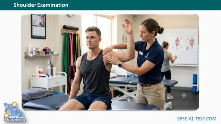
A systematic shoulder examination begins with a detailed history, followed by careful inspection, palpation, and assessment of shoulder movements. A structured approach helps identify common conditions such as rotator cuff tears, shoulder instability, adhesive capsulitis, arthritis, fractures, infections, and nerve injuries.
History Taking
Pain
Pain is the most common presenting complaint in patients with shoulder disorders. Important points to assess include:
- Onset
- Acute onset: trauma, fractures, dislocations, septic arthritis
- Insidious onset: inflammatory disorders, degenerative disease, adhesive capsulitis, tuberculosis
- Duration
- Long-standing pain is common in inflammatory arthritis, frozen shoulder, and chronic infections.
- Radiation
- Pain may radiate to:
- Posterior shoulder
- Axilla
- Lateral aspect of the upper arm
- Pain may radiate to:
- Aggravating factors
- Most painful shoulder conditions worsen with movement.
- Character of pain
- Throbbing, severe pain often suggests:
- Pyogenic infection
- Acute traumatic injury
- Throbbing, severe pain often suggests:
- Relieving factors
- Determine whether symptoms improve with:
- Rest
- Massage
- Analgesics
- Ask whether relief is complete or partial and how long it lasts.
- Determine whether symptoms improve with:
- Relationship to trauma
- Previous shoulder dislocations
- Fractures
- Fracture-dislocations
- Previous treatment
- Duration of immobilization
- Postoperative or post-injury physiotherapy
- Episodes of recurrent instability
- Pain during specific movements
- Early abduction: suggests supraspinatus tear
- Painful arc: suggests supraspinatus impingement
- Forward flexion: may indicate biceps tendon pathology
- Internal rotation (reaching behind the back): may indicate subscapularis injury
- Associated fever
- Fever raises suspicion for septic arthritis or other infective conditions.
Swelling
Determine whether swelling developed:
- Spontaneously
- Infection
- Pigmented villonodular synovitis (PVNS)
- Reactive effusion
- Inflammatory arthritis
- Hemophilia
- Degenerative disease
- Following trauma
- Hemarthrosis
- Soft tissue injury
Limitation of Movement
Ask about:
- Gradual onset
- Adhesive capsulitis
- Chronic infection
- Following treatment
- Post-traumatic stiffness
- Postoperative stiffness
Weakness (Loss of Power)
Weakness may be associated with:
- Recurrent shoulder subluxation
- Recurrent dislocation
- Dead arm syndrome
Shoulder Instability
Assess:
- Voluntary or involuntary instability
- Direction of instability
- Triggering movements
- Frequency of episodes
- Duration of symptoms
- Associated neurological symptoms or weakness
Referred Pain and Systemic Symptoms
Shoulder pain may originate outside the shoulder. Always ask about conditions involving:
- Gastric or duodenal disease
- Diaphragmatic irritation
- Cardiopulmonary disorders
- Mediastinal pathology
- Polyarthralgia suggesting systemic inflammatory disease
Inspection
Inspection should be performed from the front, back, top, and medial aspect while comparing both shoulders.
Shoulder Attitude (Posture)
Characteristic postures may indicate specific conditions:
| Clinical Finding | Possible Diagnosis |
|---|---|
| Arm abducted, externally rotated, supported by opposite hand | Anterior shoulder dislocation |
| Arm adducted and internally rotated | Posterior shoulder dislocation |
| Shoulder abducted and drooping | Deltoid contracture |
| High webbed neck | Klippel-Feil syndrome |
| Elevated scapula | Sprengel shoulder |
| Dominant scapula displaced laterally | Lateral scapular slide in throwing athletes |
| Shoulder held in flexion and abduction | Prescapular abscess |
| Mild flexion, abduction, and external rotation | Pyogenic arthritis |
Inspection from the Front
Compare both shoulders and examine:
- Sternal notch
- Sternoclavicular joint
- Clavicle and its contour
- Supraclavicular and infraclavicular fossae
- Acromioclavicular (AC) joint
- Preglenoid fossa
- Anterior axillary fold
- Coracoid prominence
- Deltoid muscle bulk
- Shoulder contour
- Pectoralis muscle
- Sternocleidomastoid muscle
- Alignment of the chin with the suprasternal notch
Inspection from Behind
Assess:
- Midline alignment
- Symmetry of the shoulders
- Trapezius muscle
- Medial border of the scapula
- Scapular winging (commonly due to serratus anterior weakness)
- Spine of the scapula
- Inferior angle of the scapula
- Supraspinous fossa
- Infraspinous fossa
- Posterior axillary fold
- Soft spot (approximately 1 cm medial and 2 cm inferior to the acromion) for swelling
Inspection from Above
Observe:
- Acromioclavicular joint
- Overall shoulder contour
Inspection from the Medial Aspect
Look for:
- Enlarged lymph nodes
- Sebaceous cysts or skin infections
Evaluate the skin using the SEADS approach:
- Swelling
- Erythema
- Atrophy
- Discoloration
- Suppuration (including scars and sinuses)
Palpation
Begin by assessing:
- Local temperature
- Superficial tenderness
Proceed to deep regional palpation.
Anterior Structures
Palpate:
- Sternoclavicular joint
- Clavicle
- Acromioclavicular joint
- Acromion (including os acromiale)
- Subacromial bursa
- Long head of the biceps tendon
- Best palpated 1–4 cm anterior to the acromion with approximately 10° of internal rotation
- Myositis masses
- Pectoralis major tendon
- Supraclavicular fossa for brachial plexus tenderness or “burners” and “stingers”
Lateral Aspect
Assess for:
- Deltoid muscle bulk
- Step deformity suggesting inferior shoulder subluxation
Posterior Aspect
Palpate the soft spot for:
- Effusion
- Swelling
- Local tenderness
Medial Aspect
Assess:
- Axillary artery pulsations
Assessment of Shoulder Movements
Both active and passive range of motion should be examined and compared with the opposite side.
Forward Flexion
- Normal range: 160–180°
Abduction
Observe:
- Scapulohumeral rhythm
- External rotation after approximately 90° of abduction
- Shoulder shrugging, which may indicate chronic rotator cuff insufficiency
Look for a painful arc, characterized by:
- Minimal pain during early abduction
- Pain during the mid-range
- Improvement near terminal abduction
This finding commonly suggests:
- Supraspinatus impingement
- Partial supraspinatus tear
Additional findings:
- Inability to initiate abduction suggests supraspinatus weakness.
- Inability to maintain abduction suggests deltoid weakness.
Adduction
Ask the patient to:
- Bring the arm across the chest
- Compare cross-body adduction with the opposite side
Internal Rotation
Assess with:
- Arm at the side (0° abduction)
- Arm abducted to 90°
Normal internal rotation at 90° abduction is approximately 45°.
The Apley Scratch Test provides a functional assessment by asking the patient to reach behind the back.
Normal findings:
- Women: thumb reaches approximately T7
- Men: thumb reaches approximately T9
External Rotation
Evaluate with:
- Arm at the side
- Shoulder abducted to 90°
Normal external rotation is approximately 90°.
Total Active Elevation
Ask the patient to elevate the arm in the plane of the scapula, approximately 20–30° anterior to the coronal plane.
Scapular Protraction
Ask the patient to:
- Round the shoulders forward
- Assume a “hunched” posture
This evaluates serratus anterior function.
Scapular Retraction
Ask the patient to:
- Pull both shoulders backward into an “attention” position
Alternating between protraction and retraction may reproduce symptoms of snapping scapula syndrome.
Clinical Pearls
- Always compare findings with the opposite shoulder.
- Observe the patient before touching the shoulder, as posture often provides important diagnostic clues.
- A painful arc strongly suggests rotator cuff pathology, particularly supraspinatus impingement.
- Fever with shoulder pain should prompt evaluation for septic arthritis or osteomyelitis.
- Shoulder pain may be referred from the neck, diaphragm, heart, lungs, or upper gastrointestinal tract, so a complete history is essential.
- Both active and passive range of motion should be documented, as preserved passive motion with restricted active motion often indicates rotator cuff pathology, whereas restriction of both suggests adhesive capsulitis or arthritis.