Shoulder Range of Motion
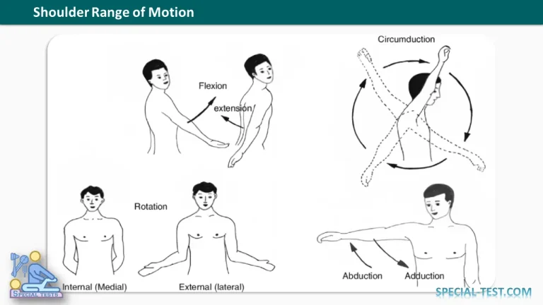
Assessment of the shoulder range of motion (ROM) is a fundamental part of every orthopedic and musculoskeletal examination. Measuring active and passive movements helps identify pathology involving the glenohumeral joint, acromioclavicular joint, sternoclavicular joint, scapulothoracic articulation, rotator cuff, and surrounding soft tissues.
A reduction in shoulder ROM may result from pain, muscle weakness, tendon injury, joint stiffness, adhesive capsulitis, arthritis, fractures, or neurological disorders. Comparing the affected shoulder with the contralateral side is essential because slight variations exist among healthy individuals.
Normal Shoulder Range of Motion
The following table summarizes the normal range of motion of the shoulder.
| Movement | Normal Range of Motion (Degrees) |
|---|---|
| Abduction | 0–170° |
| Adduction | 0–50° |
| Forward Flexion | 0–165° |
| Backward Extension | 0–60° |
| Internal Rotation | Approximately 70° |
| External Rotation | 70–100° |
Note: Normal values vary slightly among references depending on patient age, sex, flexibility, and the measurement technique used.
How to Measure Shoulder Range of Motion
Shoulder ROM should be assessed in both active and passive movements.
- Active ROM evaluates muscle strength, tendon integrity, coordination, and pain.
- Passive ROM assesses the joint capsule, ligaments, and articular surfaces while minimizing muscular contribution.
A goniometer is the standard instrument used for accurate measurement in clinical practice.
Individual Shoulder Movements
Shoulder Flexion
Forward flexion moves the arm anteriorly in the sagittal plane.
- Normal ROM: 0–165° to 180°
- Primary muscles:
- Anterior deltoid
- Clavicular head of pectoralis major
- Coracobrachialis
- Biceps brachii (long head)
Painful limitation may indicate:
- Rotator cuff disease
- Subacromial impingement
- Glenohumeral arthritis
- Adhesive capsulitis
Shoulder Extension
Extension moves the arm posteriorly.
- Normal ROM: 0–60°
- Primary muscles:
- Posterior deltoid
- Latissimus dorsi
- Teres major
- Long head of triceps
Reduced extension may occur following trauma, shoulder stiffness, or degenerative joint disease.
Shoulder Abduction
Abduction is raising the arm away from the body in the coronal plane.
- Normal ROM: 0–170° to 180°
The movement occurs in phases:
- 0–15°: Supraspinatus initiates movement.
- 15–90°: Middle deltoid becomes the primary abductor.
- Above 90°: Scapular upward rotation is required.
Normal abduction depends on the coordinated movement of the glenohumeral and scapulothoracic joints, known as the scapulohumeral rhythm.
Shoulder Adduction
Adduction brings the arm toward the trunk.
- Normal ROM: 0–50°
Primary muscles include:
- Pectoralis major
- Latissimus dorsi
- Teres major
Internal Rotation
Internal (medial) rotation rotates the humerus toward the body.
- Normal ROM: Approximately 70°
Primary muscles:
- Subscapularis
- Pectoralis major
- Latissimus dorsi
- Teres major
- Anterior deltoid
Clinically, internal rotation is often assessed by asking the patient to place the thumb behind the back and noting the highest vertebral level reached.
External Rotation
External (lateral) rotation rotates the humerus away from the body.
- Normal ROM: 70–100°
Primary muscles:
- Infraspinatus
- Teres minor
- Posterior deltoid
Loss of external rotation is one of the earliest findings in adhesive capsulitis (frozen shoulder).
Scapulohumeral Rhythm
Normal shoulder motion depends on coordinated movement between the glenohumeral joint and the scapula.
During full arm elevation:
- Approximately 120° occurs at the glenohumeral joint.
- Approximately 60° results from scapular upward rotation.
This produces the classic 2:1 scapulohumeral rhythm, meaning that for every 3 degrees of shoulder elevation, about 2 degrees occur at the glenohumeral joint and 1 degree occurs through scapular rotation.
Disruption of this rhythm may indicate:
- Rotator cuff tears
- Scapular dyskinesis
- Long thoracic nerve palsy
- Shoulder impingement syndrome
Clinical Significance
Restriction of shoulder ROM provides valuable diagnostic clues.
| Limited Movement | Possible Causes |
|---|---|
| Painful abduction | Rotator cuff tendinopathy, impingement syndrome, calcific tendinitis |
| Global restriction of active and passive ROM | Adhesive capsulitis |
| Limited passive ROM with crepitus | Glenohumeral osteoarthritis |
| Weak active ROM with preserved passive ROM | Rotator cuff tear or neurological injury |
| Painful external rotation | Infraspinatus or teres minor pathology |
| Limited internal rotation | Posterior capsule tightness, glenohumeral internal rotation deficit (GIRD), adhesive capsulitis |
Because many shoulder disorders present with overlapping symptoms, ROM assessment should always be combined with inspection, palpation, muscle strength testing, and special orthopedic tests.
Factors Affecting Shoulder Range of Motion
Several factors influence normal shoulder mobility, including:
- Age
- Sex
- Hand dominance
- Athletic participation
- Previous injuries or surgery
- Pain
- Muscle tightness
- Capsular contracture
- Neurological disorders
Older adults generally have slightly reduced ROM compared with younger individuals due to age-related changes in connective tissue elasticity.
Key Points
- Shoulder ROM should be measured bilaterally using both active and passive movements.
- A goniometer provides the most accurate clinical measurements.
- Normal shoulder elevation requires coordinated glenohumeral and scapulothoracic motion.
- Loss of external rotation is an early hallmark of adhesive capsulitis.
- Comparing active and passive ROM helps distinguish muscular pathology from joint stiffness.
References
- Almoallim H, Kalantan D, Alharbi L, Albazli K. Approach to Musculoskeletal Examination. In: Skills in Rheumatology. Springer; 2021.
- Magee DJ. Orthopedic Physical Assessment. 7th Edition. Elsevier.
- American Academy of Orthopaedic Surgeons (AAOS). Shoulder Examination and Range of Motion.
- Norkin CC, White DJ. Measurement of Joint Motion: A Guide to Goniometry. 5th Edition.
- Clinical Methods: The History, Physical, and Laboratory Examinations. 3rd Edition. National Center for Biotechnology Information (NCBI Bookshelf).
- StatPearls. Shoulder Examination and Shoulder Anatomy. StatPearls Publishing, NCBI Bookshelf.