Deltoid Ligament Test: Steps, Purpose & Positive Signs
Here is an SEO-optimized version of your medical examination guide. It is structured with a clear keyword-rich title, H2/H3 headings for search engines and screen readers, bullet points for readability, and optimized meta-data to improve click-through rates (CTR) and rankings.
Deltoid Ligament Test: Purpose, Procedure, & Positive Results
Quick Summary: The Deltoid Ligament Test is a specialized orthopedic assessment used to evaluate the integrity of the deltoid ligament complex in the ankle. It uses three distinct passive movements to target the anterior, middle, and posterior fibers.
What is the Deltoid Ligament Test?
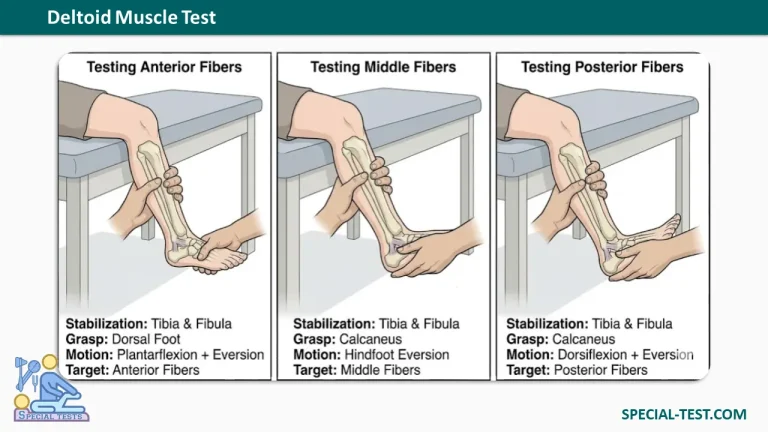
The Deltoid Ligament Test is a clinical orthopedic examination used to assess the stability and integrity of the deltoid ligament (the strong medial ligamentous complex of the ankle). The test isolates the ligament’s distinct anatomical bands by applying three separate passive movements combining eversion with varying degrees of ankle flexion.
Patient Positioning & Setup
- Patient Position: Seated comfortably with the leg flexed at the knee and the lower leg hanging freely over the edge of the treatment table.
- Examiner Position: Seated or standing facing the patient.
- Stabilization: Use one hand to firmly stabilize the anterior surface of the tibia and fibula immediately proximal to the ankle joint.
Step-by-Step Procedure
To perform a thorough evaluation, assess each fiber bundle sequentially using the free hand:
1. Assessing the Anterior Fibers
- Action: Grasp the dorsal surface of the foot with your mobilizing hand.
- Movement: Passively combine eversion and plantarflexion of the foot.
- Overpressure: Apply gentle, firm overpressure at the end range.
2. Assessing the Middle Fibers
- Action: Reposition your mobilizing hand to securely grasp the calcaneus (hindfoot).
- Movement: Passively take the patient’s hindfoot into eversion in a neutral position.
- Overpressure: Apply gentle overpressure at the end range.
3. Assessing the Posterior Fibers
- Action: Maintain your grasp on the calcaneus (hindfoot).
- Movement: Passively combine eversion and dorsiflexion of the foot.
- Overpressure: Apply gentle overpressure at the end range.
Interpreting Test Results
- Positive Sign: Pain localized to the medial ankle over the deltoid ligament, accompanied by hypermobility compared to the uninjured side.
- End Feel Note: A muscle spasm end-feel may be noted if the patient presents with a subacute injury.
References
- Al-Mohrej, Omar & Al-Kenani, Nader. (2016). Acute ankle sprain: Conservative or surgical approach? EFORT Open Reviews, 1, 28-38.
- Clinical Tests for the Musculoskeletal System, 3rd Edition.
- Dutton’s Orthopaedic Examination, Evaluation, And Intervention, 3rd Edition.