Photostress Test: Procedure, Normal Values and Interpretation
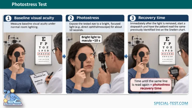
The Photostress Test, also known as the photostress recovery test (PSRT), is a noninvasive ophthalmological examination used to assess macular function by measuring the time required for visual recovery following exposure to bright light.
The test is particularly useful as an additional clinical examination when differentiating visual impairment caused by macular disorders from visual loss associated with optic nerve dysfunction.
Although modern diagnostic imaging techniques provide more detailed information about retinal abnormalities, the photostress recovery test remains a useful supplementary tool in ophthalmology and neuro-ophthalmology.
What Is the Photostress Test?
The photostress test is a clinical examination that evaluates the ability of the macula to recover its visual function following temporary exposure to a controlled bright light source.
The macula is a specialized area near the center of the retina responsible for central vision, color perception, reading, facial recognition and other activities requiring fine visual detail.
Following exposure to bright light, retinal sensitivity temporarily decreases. In healthy eyes, visual function gradually returns toward its previous level. However, patients with certain macular diseases may experience a significantly longer recovery period.
The duration required for visual recovery is known as the photostress recovery time (PRT).
Purpose of the Photostress Test
The primary purpose of the photostress test is to evaluate macular function and assist in differentiating retinal abnormalities from optic nerve disorders.
Clinical applications of the test include:
- Assessment of unexplained central visual loss.
- Evaluation of suspected macular dysfunction.
- Differentiation between macular disease and optic neuropathy.
- Assessment of visual recovery following glare exposure.
- Supplementary evaluation of age-related macular degeneration (AMD).
- Investigation of asymmetric visual impairment between the two eyes.
- Research into changes in macular function over time.
The photostress test should not be used as a standalone diagnostic examination. Its findings must be interpreted alongside other ophthalmological investigations.
Physiological Basis of the Photostress Test
The photostress recovery test is based on the physiological processes of retinal photopigment bleaching and regeneration.
When the retina is exposed to bright light, light-sensitive visual pigments within the photoreceptor cells undergo temporary bleaching. This process reduces retinal sensitivity and may produce a temporary afterimage.
After the light source is removed, the visual pigments regenerate through processes involving the photoreceptors and the retinal pigment epithelium (RPE).
In individuals with healthy macular function, visual sensitivity generally recovers relatively quickly. However, diseases affecting the macular photoreceptors or retinal pigment epithelium may prolong this recovery process.
Because certain optic nerve disorders do not directly disrupt retinal photopigment regeneration, photostress recovery may remain relatively normal despite reduced visual acuity.
This physiological difference forms the basis for the clinical application of the photostress test. [1,2]
How Is the Photostress Test Performed?
The photostress recovery test should be performed by a trained healthcare professional using appropriate ophthalmological equipment.
Patient Preparation
The patient is positioned comfortably in front of a standardized visual acuity chart. Best-corrected visual acuity is measured separately in each eye before testing.
The examiner explains that temporary glare, blurred vision or an afterimage may occur following light exposure.
Photostress Test Procedure
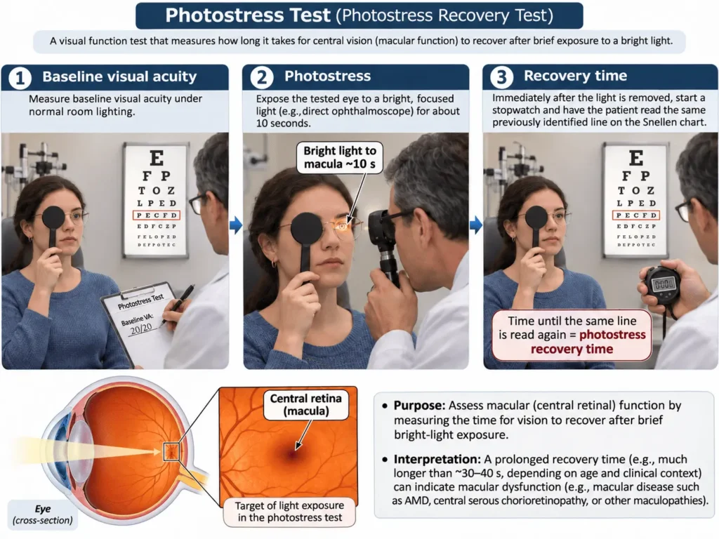
- Measure and record the patient’s baseline visual acuity.
- Cover the eye that is not being examined.
- Ask the patient to maintain fixation as directed by the examiner.
- Expose the macular region to an appropriate, controlled ophthalmic light source for the duration specified by the selected protocol. Traditional clinical methods have used approximately 10 seconds of exposure.
- Remove the light source and immediately start a stopwatch.
- Ask the patient to read the predetermined line on the visual acuity chart.
- Record the time required to recover to the specified visual acuity endpoint.
- Repeat the procedure on the opposite eye under equivalent testing conditions.
Some standardized research protocols use a 30-second exposure with a direct ophthalmoscope. These protocols may produce different recovery times from traditional penlight testing. [1,3]
Photostress Test Normal Values
Normal photostress recovery time varies depending on the patient’s age, the examination technique, the light source, exposure duration and the definition of visual recovery.
In traditional clinical assessments, recovery often occurs within several tens of seconds. The NCBI Bookshelf clinical review describes recovery exceeding 50 seconds as abnormal within its clinical testing framework.
However, this threshold is not universally applicable.
| Photostress test finding | Clinical interpretation |
|---|---|
| Rapid and symmetrical recovery | Generally consistent with preserved macular recovery function |
| Recovery exceeding 50 seconds | Potentially abnormal under certain traditional clinical protocols |
| Significantly prolonged recovery | May suggest macular dysfunction |
| Delayed recovery in one eye | May indicate asymmetric macular impairment |
| Normal recovery despite reduced visual acuity | May support investigation of an optic nerve disorder |
Longer recovery times may occur in healthy older adults, particularly when more intensive photostress protocols are used. Therefore, age-appropriate and protocol-specific reference values are essential. [3,4,5]
What Is a Positive Photostress Test?
A positive photostress test refers to an abnormally prolonged recovery of visual function following controlled light exposure.
An abnormal result may be associated with conditions affecting the macula, including:
- Age-related macular degeneration.
- Diabetic macular disease.
- Macular edema.
- Retinal pigment epithelium abnormalities.
- Other disorders affecting central retinal photoreceptors.
A significant difference in recovery time between the two eyes may also raise suspicion of unilateral or asymmetric macular pathology.
Nevertheless, a positive photostress test does not establish a definitive diagnosis or identify a specific underlying disease. Additional ophthalmological examinations are necessary.
Photostress Test in Age-Related Macular Degeneration
Age-related macular degeneration (AMD) is one of the most extensively studied conditions associated with prolonged photostress recovery.
A study published in the journal Ophthalmology found delayed recovery in 62% of the studied patients with age-related macular degeneration. Delayed recovery was also observed in 47% of participants who had normal visual acuity. [6]
A population-based study published in 2023 reported an association between prolonged photostress recovery time and both early and advanced AMD in older adults. The findings suggest that photostress recovery may have potential as a functional biomarker for macular disease. [5]
However, further research and standardization are required before photostress testing can be relied upon as an independent screening or predictive tool.
Photostress Test: Macular Disease vs. Optic Neuropathy
One of the classical applications of the photostress recovery test is distinguishing macular disease from optic nerve dysfunction.
| Clinical feature | Macular disease | Optic neuropathy |
|---|---|---|
| Visual acuity | Normal or reduced | Normal or reduced |
| Photostress recovery time | Often prolonged | May remain relatively normal |
| Visual distortion | May be present | Less characteristic |
| Primary affected structure | Central retina | Optic nerve |
These differences are general clinical patterns rather than definitive diagnostic criteria.
Additional examinations, including pupillary assessment, color vision testing, visual field testing, fundus examination and optical coherence tomography (OCT), may be necessary to determine the cause of visual impairment.
Advantages and Limitations
The photostress recovery test is relatively inexpensive, noninvasive and simple to perform in a clinical setting. It provides functional information about the macula that may complement anatomical findings.
However, several factors can influence its accuracy and reliability:
- Patient age.
- Light intensity and exposure duration.
- Differences in examination protocols.
- Poor visual fixation.
- Difficulty reading the visual acuity chart.
- Coexisting ocular abnormalities.
- Patient cooperation and attention.
- Variability between repeated measurements.
Research has also demonstrated that certain photostress testing devices have limited usefulness in assessing the severity or progression of macular degeneration. [7]
Consequently, a normal test result does not exclude retinal disease, and an abnormal result does not necessarily confirm macular pathology.
Frequently Asked Questions (FAQ)
What does the photostress test measure?
The photostress test measures the time required for central visual function to return toward its baseline level following controlled exposure to bright light.
What is the normal photostress recovery time?
Normal recovery generally occurs within several tens of seconds using traditional clinical methods. However, reference values vary according to age, light exposure and the testing protocol.
What does a positive photostress test indicate?
A positive test indicates delayed visual recovery, which may suggest macular dysfunction. Further examinations are required to establish the underlying cause.
Can the photostress test detect macular degeneration?
The test may identify functional changes associated with age-related macular degeneration, including changes in some patients with normal visual acuity. However, it cannot independently diagnose or exclude AMD.
Is the photostress test painful?
The examination is generally noninvasive and painless. Some individuals may experience temporary glare, visual discomfort or an afterimage.
Can I perform a photostress test at home?
Home photostress testing with improvised light sources is not recommended. The examination should be performed by a trained eye-care professional using suitable equipment and standardized techniques.
Can the photostress test replace optical coherence tomography (OCT)?
No. Photostress testing evaluates functional visual recovery, whereas OCT provides detailed anatomical images of the retina. These examinations provide different types of clinical information.
Does age affect photostress recovery time?
Yes. Research demonstrates that visual recovery following photostress may become slower with increasing age, even in individuals without identifiable retinal disease.
Can a normal photostress test rule out optic nerve disease?
No. Patients with certain optic nerve disorders may have normal photostress recovery despite impaired visual function.
Conclusion
The photostress test is a supplementary ophthalmological examination used to evaluate macular function and assist in differentiating macular disorders from optic nerve diseases.
Although prolonged photostress recovery may indicate retinal dysfunction, the results should always be interpreted within the context of the patient’s medical history, clinical examination and appropriate diagnostic imaging.
Patients experiencing sudden visual loss, new central visual distortion or a dark spot in their central vision should seek urgent ophthalmological assessment.
References
- Glaser JS, Savino PJ, Sumers KD, et al. The photostress recovery test in the clinical assessment of visual function. American Journal of Ophthalmology. 1977. PubMed
- Macular photostress testing: sensitivity and recovery with an automated perimeter. PubMed
- Sources of variability in the clinical photostress test. PubMed
- Tests for Potential Vision. StatPearls. NCBI Bookshelf
- Photostress Recovery Time as a Potential Predictive Biomarker for Age-Related Macular Degeneration. 2023. PubMed Central
- Slow photostress recovery and disease severity in age-related macular degeneration. PubMed
- Effect of age related macular degeneration on the Eger macular stressometer photostress recovery time. PubMed Central
- The macular automated photostress test. PubMed
- Age-related macular degeneration: diagnosis and management. NICE Guideline No. 82. NCBI Bookshelf